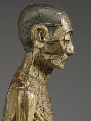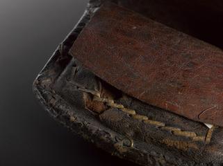
Papier mache anatomical figure, covered with human skin, Japanese
This small Japanese papier-mâché model of the body has the chest cut away to reveal the ribs, lungs, heart and arteries. Its teeth and tongue are visible, and the figure is wrapped in preserved human skin.
Before the invention of the X-ray machine in 1895, seeing inside the human body was restricted to the dissection theatre. However, this scope was limited. Most cultures had religious or ethical taboos about dissection. Cadavers were also difficult to acquire and preserve. Anatomical models allow students to study the body’s internal structures in detail without the need for a dissection.
Details
- Category:
- Asian Medicine
- Collection:
- Sir Henry Wellcome's Museum Collection
- Object Number:
- A661119
- Measurements:
-
overall: 57 mm x 110 mm x 410 mm, .394kg
- type:
- human remains, human skin and anatomical figure
- credit:
- Wellcome Trust


