
Wet plate negative copper spark spectrum by Dr. Miller
![]() This image is released under a CC BY-NC-SA 4.0 Licence
This image is released under a CC BY-NC-SA 4.0 Licence
Buy this image as a print
BuyLicense this image for commercial use at Science and Society Picture Library
LicenseWet plate negative copper spark spectrum by Dr. Miller
Science Museum Group
© The Board of Trustees of the Science Museum
Wet plate negative copper spark spectrum by Dr. Miller
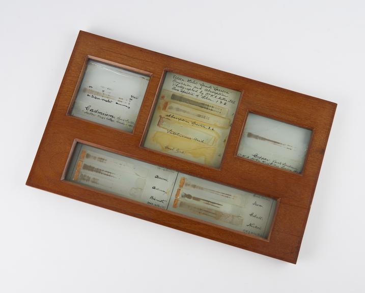
In 1845, William Allen Miller (professor of chemistry at King’s College London) hand drew the spectra of calcium, copper and barium chloides, boric acid and strontium nitrate. He noticed that each displayed several bright lines and bands, and all having the bright yellow ‘D line’ (a feature of the spectra of sodium compounds) in common.
In 1862, Miller studies spectra of a number of metals into the ultra-violet region. Instead of examining them on a fluorescent screen, he photographed his spectra, commenting:
“The lines of each spectrum are so numerous and so close together that it would be impossible without sacrifice of time, that would scarely be justifiable, to obtain accurate impressions of them by eye-drawing. Indeed, except by the process of photography, these lines can only be rendered visible by the air of a fluorescent screen, under which circumstances the minute details are almost necessarily lost even by the most careful observer.”
These plates are the earliest surviving photographs of ultra-violet spectra. Miller used a wet collodion plate, complaining of the comparatively low dispersion of quartz and of the disadvantage of the effects of double refraction. The plates show spark spectra of iron, cobalt, nickel, arsenic, antimony, bismuth, copper and silver. Additionally, there are two absorption spectra of sulphurous acid (almost invisible) and coal gas (only fragments remaining).
Details
- Category:
- Experimental Chemistry
- Object Number:
- 1914-380
- type:
- ultraviolet spectra
- credit:
- Hartley, W.J.


