![]() This image is released under a CC BY-NC-SA 4.0 Licence
This image is released under a CC BY-NC-SA 4.0 Licence
Buy this image as a print
BuyLicense this image for commercial use at Science and Society Picture Library
License![]() This image is released under a CC BY-NC-SA 4.0 Licence
This image is released under a CC BY-NC-SA 4.0 Licence
Buy this image as a print
BuyLicense this image for commercial use at Science and Society Picture Library
License![]() This image is released under a CC BY-NC-SA 4.0 Licence
This image is released under a CC BY-NC-SA 4.0 Licence
Buy this image as a print
BuyLicense this image for commercial use at Science and Society Picture Library
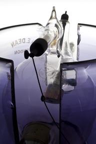
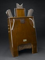
License"Tomograph" x-ray machine with couch, by Sanitas of Berlin
Science Museum Group Collection
© The Board of Trustees of the Science Museum
"Tomograph" x-ray machine with couch, by Sanitas of Berlin
Science Museum Group Collection
© The Board of Trustees of the Science Museum
"Tomograph" x-ray machine with couch, by Sanitas of Berlin
Science Museum Group Collection
© The Board of Trustees of the Science Museum
Tomography x-ray machine with couch, used at Preston Hall Hospital, Kent, made by Sanitas, Freidrichstrasse Bld., Berlin, Germany, 1933-1940.
X-ray machines are used by radiographers to acquire an image of the body’s skeletal structure. X-rays can also be used to detect disease or abnormalities in soft tissue such as the chest. A tomography machine creates X-ray images of sections or slices of the body, known as tomograms. This was a new technique in the 1930s. It diagnosed diseases such as pulmonary tuberculosis more effectively.
The use of tomography in England was pioneered by Dr J B McDougall at Preston Hall Hospital in Kent. This example was presented to the Wellcome collections by the hospital in 1964.
Details
- Category:
- Radiomedicine
- Collection:
- Sir Henry Wellcome's Museum Collection
- Object Number:
- A639432
- Materials:
- metal
- Measurements:
-
overall: 2000 mm x 1350 mm x 1300 mm,
- type:
- x-ray machine
- credit:
- Preston Hall Hospital