


![]() This image is released under a CC BY-NC-SA 4.0
Licence
This image is released under a CC BY-NC-SA 4.0
Licence
Buy this image as a print
BuyLicense this image for commercial use at Science and Society Picture Library
License![]() This image is released under a CC BY-NC-SA 4.0
Licence
This image is released under a CC BY-NC-SA 4.0
Licence
Buy this image as a print
BuyLicense this image for commercial use at Science and Society Picture Library
LicensePhotograph of an early method of testing the output of X-rays
Science Museum Group
© The Board of Trustees of the Science Museum
Science Museum Group Collection
© The Board of Trustees of the Science Museum
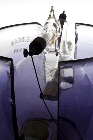
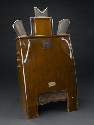
Photograph of an early method of testing the output of X-rays by observing the appearance of a hand through a fluoroscope
An early method of testing the output of X-rays using a fluoroscope is shown in this photograph. The body could be viewed through a fluoroscope without taking or developing time-consuming X-ray photographs. The X-rays instead fell on a screen which fluoresced immediately. This let the physician see the same thing as an X-ray photograph, or in this case use the fluoroscope to test the X-ray output.
X-rays were discovered in 1895 by German physicist Wilhelm Röntgen (1845-1923). Battlefield surgeons and medics quickly took them up. However, X-ray departments were rare in hospitals in the UK until the 1930s. This was because of the costs and the need for trained personnel. Early X-rays also took time to develop. The images were sometimes poor quality.
Details
- Category:
- Radiomedicine
- Collection:
- Sir Henry Wellcome's Museum Collection
- Object Number:
- A606970
- Materials:
- paper and cover, glass
- type:
- photograph