
![]() This image is released under a CC BY-NC-SA 4.0
Licence
This image is released under a CC BY-NC-SA 4.0
Licence
Buy this image as a print
BuyLicense this image for commercial use at Science and Society Picture Library
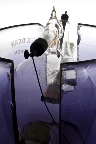
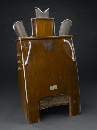
LicenseDouble focus X-ray tube, 1896. Graduated white background.
Science Museum Group Collection
© The Board of Trustees of the Science Museum
Double focus X-ray tube, 1896.
This tube worked by using an alternating electric current, which accelerates electrons towards an aluminium plate. This produced x-rays at both ends of the tube. Wilhelm Röntgen (1845-1923), a German physician, took the first x-ray in 1896 of his wife’s left hand. Dense areas of bone show up as white whilst soft tissue allow the x-ray to pass through undeterred. Very quickly x-rays proved their usefulness as a diagnostic and therapeutic tool in medicine. Within six months of Röntgen’s announcement x-rays were being used by battlefield physicians to locate bullets in wounded soldiers. X-rays allowed physicians their first look inside the body without resorting to surgery.
Details
- Category:
- Radiomedicine
- Collection:
- Sir Henry Wellcome's Museum Collection
- Object Number:
- A201
- Materials:
- wood, glass, aluminium (cathodes) and platinum (anticathodes)
- Measurements:
-
overall: 210 mm x 270 mm x 77 mm,
diameter (sphere): 75 mm,
diameter (stand base): 77 mm,
diameter (arm): 25 mm,
- type:
- x-ray tube
- credit:
- Wellcome Trust, purchased from F Holmes