


![]() This image is released under a CC BY-NC-SA 4.0
Licence
This image is released under a CC BY-NC-SA 4.0
Licence
Buy this image as a print
BuyLicense this image for commercial use at Science and Society Picture Library
License![]() This image is released under a CC BY-NC-SA 4.0
Licence
This image is released under a CC BY-NC-SA 4.0
Licence
Buy this image as a print
BuyLicense this image for commercial use at Science and Society Picture Library
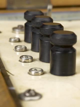
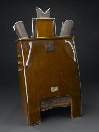
LicensePotter Bucky diaphragm, sectioned, English, 1940-1950
Science Museum Group
© The Board of Trustees of the Science Museum
Potter Bucky diaphragm, sectioned, English, 1940-1950
Science Museum Group Collection
© The Board of Trustees of the Science Museum
Potter Bucky diaphragm, sectioned, English, 1940-1950
Early X-ray plates were blurred due to the scattering of rays by body tissue. Radiologists Gustav Bucky (1880–1963) and Hollis E. Potter (1880-1964) worked independently on this problem from about 1913. A section of the resultant Bucky-Potter Diaphragm is shown. It was placed between the patient and the X-ray film. Parallel lead strips move across inside the wooden casing during an exposure. They block scattered rays travelling at other angles. The lead strips are not visible on the X-ray film because they are moving. The device was first marketed in 1921. It has become standard equipment on X-ray tables.
Details
- Category:
- Radiomedicine
- Object Number:
- 1980-1221
- Measurements:
-
overall: 120 mm x 590 mm x 350 mm,
- type:
- x-ray accessory
- credit:
- Hypher, T.J.