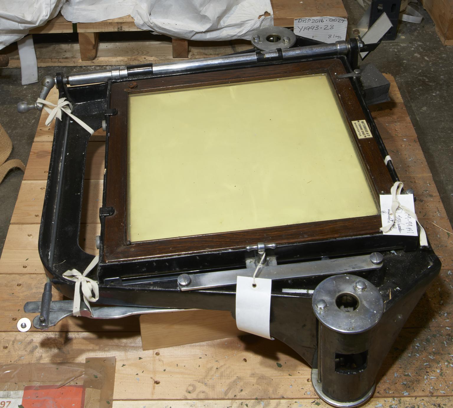
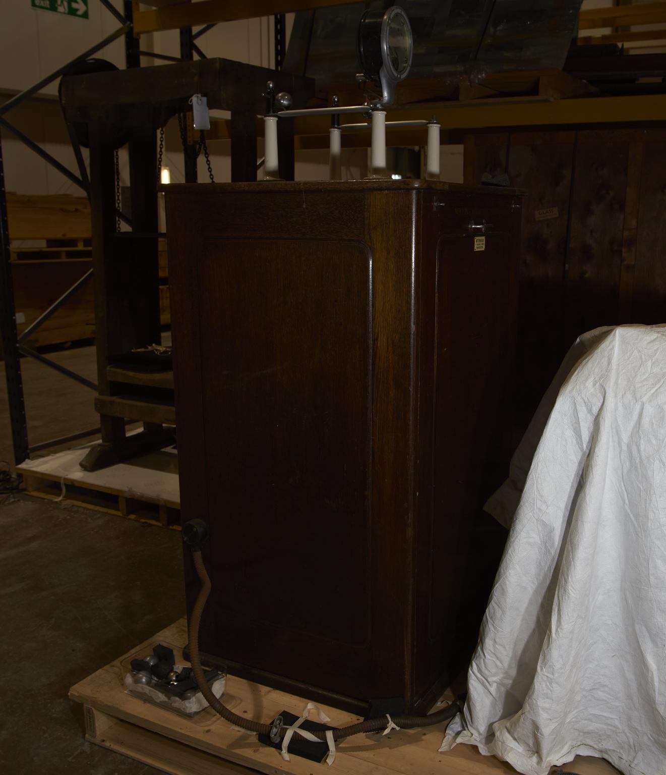
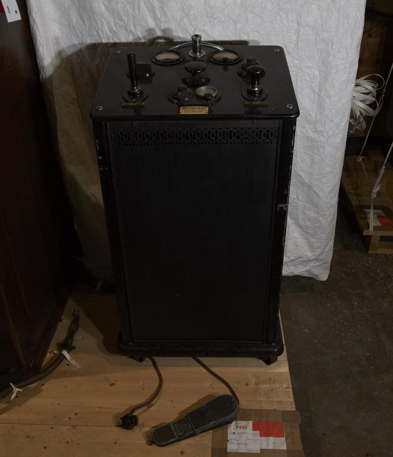
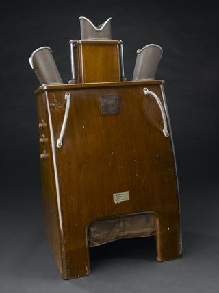
X-ray set comprising fluoroscopic chest screening frame, couch with facility for fluoroscopy and x-ray photography, protective apron, power supply and control unit, by Newton and Wright Limited, London, England, 1920-1923, used until 1960.
Fluoroscopy allows X-rays to be viewed without taking and developing X-ray photographs. This X-ray machine doubles as a fluoroscopic chest screening frame. It is shown in a reconstruction of a 1930s X-ray room. The set also includes a control panel to set the dosage levels, and a screen to protect the radiographer. X-rays were discovered in 1895 by German physicist Wilhelm Röntgen (1845-1923). They allowed physicians their first look inside the body without surgery.
This X-ray machine was originally used at Westmorland County Hospital in Kendal, Cumbria, England. It was transferred to the private surgery in a doctor’s home in the 1940s. The machine was made by Newton & Wright Limited. It remained in use until the 1960s. It was probably quite unusual that physicians owned their own equipment. This was because the machines were expensive to buy and maintain. X-ray departments were rare in hospitals before the First World War. However, almost every hospital in the UK had one by the 1930s.
