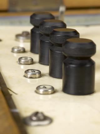
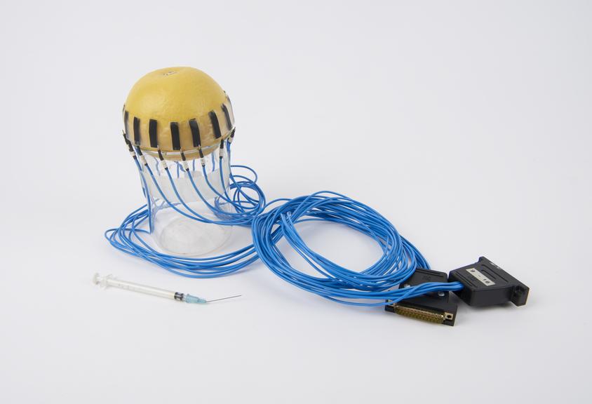
Set of 16 prototype electrodes for applied potential tomography (APT) body imaging, 1987.
- Made:
- 1987
![]() This image is released under a CC BY-NC-SA 4.0
Licence
This image is released under a CC BY-NC-SA 4.0
Licence
Buy this image as a print
BuyLicense this image for commercial use at Science and Society Picture Library
LicenseSet of 16 electrodes for APT (Applied Potential Tomography)
Science Museum Group
© The Board of Trustees of the Science Museum
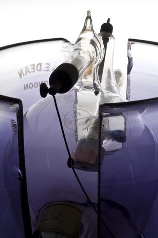
Set of 16 electrodes for APT (Applied Potential Tomography) body imaging, connected to wires terminating in two D-plugs, unsigned, English, 1987. Mounted in glass jar on artificial plastic fruit, with hypodermic syringe (needle inserted).
The electrodes are illustrated attached to a grapefruit which is standing in for a child's head and liquid is injected into the 'head' to mimic a brain haemorrhage.
Details
- Category:
- Radiomedicine
- Object Number:
- 1987-387
- Materials:
- rubber (unidentified), plastic (unidentified), metal (unknown), electronic components, adhesive tape and glass
- Measurements:
-
overall: 240 mm x 270 mm x 230 mm, .54kg
- type:
- component - object, x-ray, electrode and hypodermic syringe
- credit:
- Royal Hallamshire Hospital. Dept. of Med