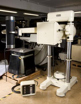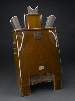
Mallard MRI body scanner, Aberdeen, Scotland, 1983
- maker:
- M&D Technology Limited


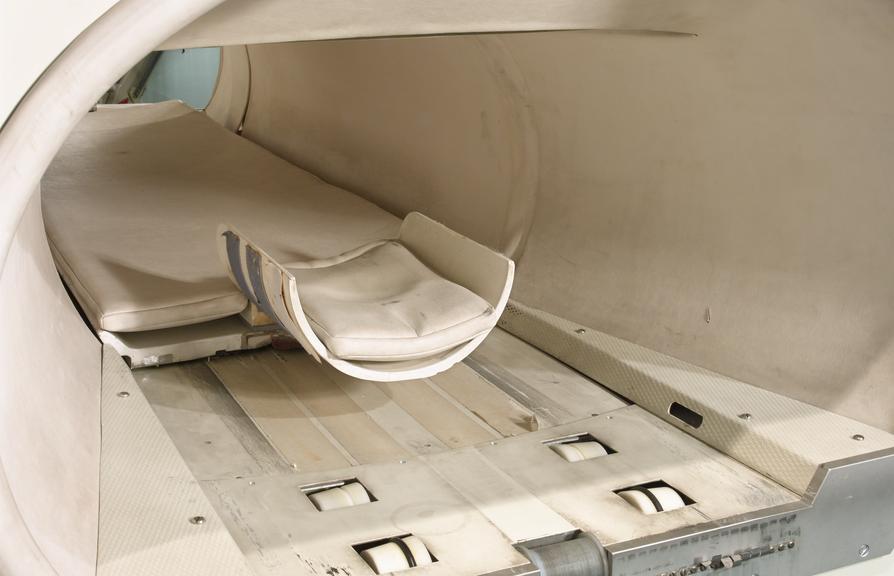
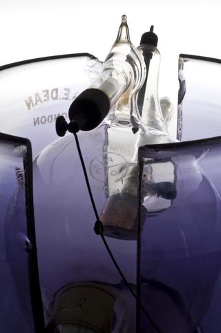
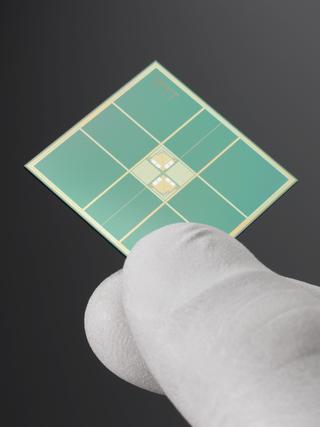
Mallard system Magnetic Resonance Imager (MRI) body scanner by M&D Technology, Aberdeen, installed at St Bartholomew's Hospital in 1983 and 1994-492 used clinically until 1993. One of three commercially-produced second-generation MRI machines manufactured by this company in Britain.
John Mallard and his research team at Aberdeen Royal Infirmary used a machine like this in 1980 when they obtained the first clinically useful image of a patient’s internal tissues using Magnetic Resonance Imaging (MRI). Mallard’s team, based at the University of Aberdeen, was responsible for technological advances that led to the widespread introduction of MRI.
MRI is considered to be a safer diagnostic tool than X-rays and is more suitable for soft tissue. MRI builds up a picture of the human body by using high frequency radio signals. Made by M&D Technology Ltd, a company set up by John Mallard, this machine was used at St Bartholomew’s Hospital, London, from 1983 to 1993.
Details
- Category:
- Radiomedicine
- Object Number:
- 1994-492
- type:
- mri body scanner
- credit:
- St Bartholomew's Hospital
