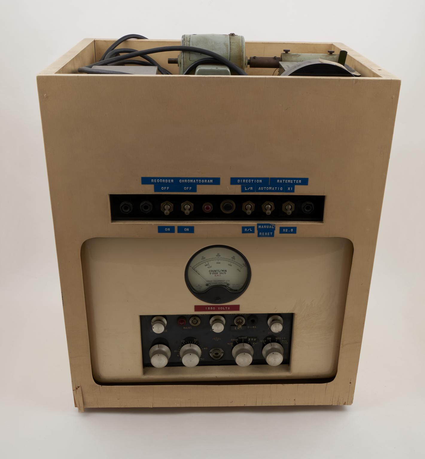
Part of automatic recording scanner for radio chromatograms, 1958
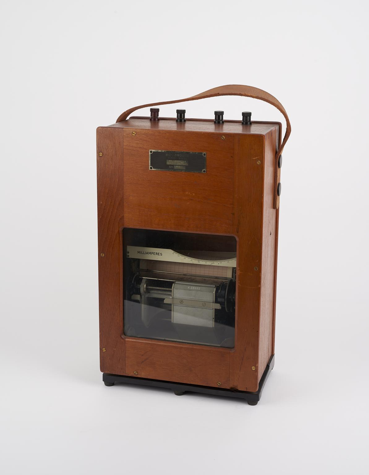
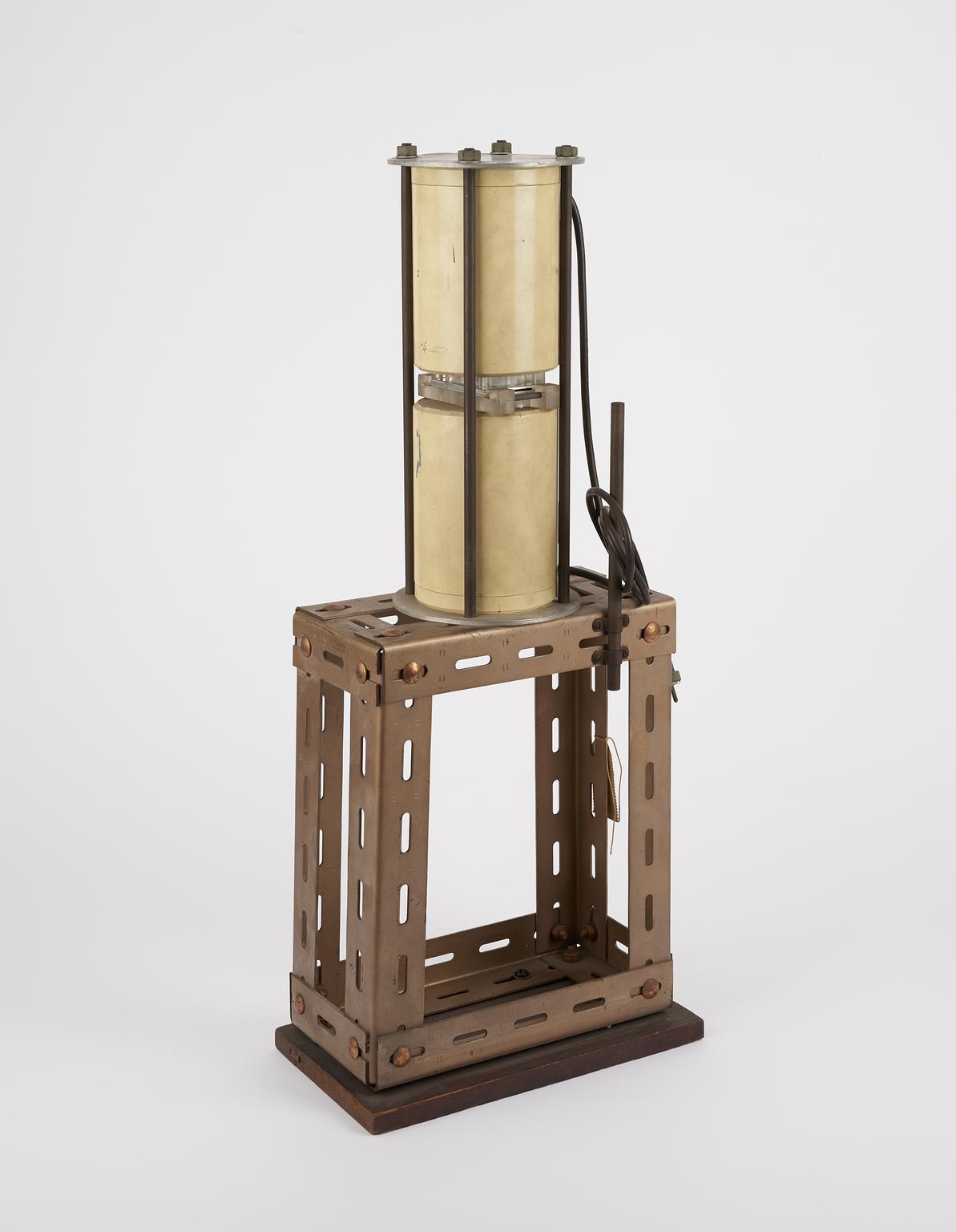
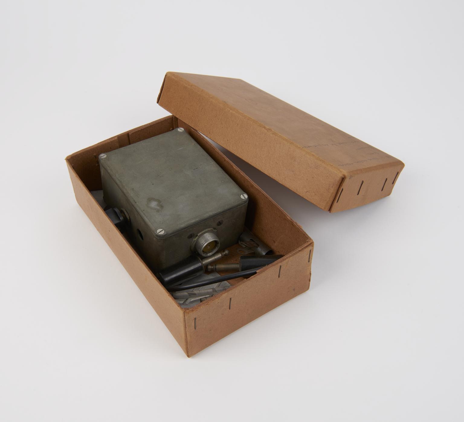
Part (unidentified) of automatic recording scanner for radio chromatograms, 1958
More
Radio chromatography is a technique used to separate and analyse components of a mixture, particularly when those components are radioactive or labelled with radioactive isotopes.
Radioactive compounds were detected by passing strip chromatograms through the automatic recording scanner. The chromatograms were scanned automatically and approximately assessed the amount of radioactivity in the sample, with the output graphically recorded by the pen-recording milliammeter.
This is part of an apparatus used in the late 1950s at the Department of Biochemistry in St Thomas's Hospital Medical School, London, by biochemists Professor Leslie Young (1911-1992) and A.R. Morrison (dates unknown). Young spent much of his career studying toxic compounds and their impacts on molecules in the body, and this equipment was used in this research. They described the equipment in the Biochemical Journal in 1959 in the article: An Automatic Recording Scanner for Radiochromatograms.
- Object Number:
- 1982-542/1
- type:
- part and radio chromatogram recording scanner
- Image ©
- The Board of Trustees of the Science Museum