


![]() This image is released under a CC BY-NC-SA 4.0
Licence
This image is released under a CC BY-NC-SA 4.0
Licence
License this image for commercial use at Science and Society Picture Library
License![]() This image is released under a CC BY-NC-SA 4.0
Licence
This image is released under a CC BY-NC-SA 4.0
Licence
License this image for commercial use at Science and Society Picture Library
License![]() This image is released under a CC BY-NC-SA 4.0
Licence
This image is released under a CC BY-NC-SA 4.0
Licence
License this image for commercial use at Science and Society Picture Library
LicenseEye defect teaching model, on wood stand, probably English
Science Museum Group Collection
© The Board of Trustees of the Science Museum
Eye defect teaching model, on wood stand, probably English
Science Museum Group Collection
© The Board of Trustees of the Science Museum
Eye defect teaching model, on wood stand, probably English
Science Museum Group Collection
© The Board of Trustees of the Science Museum
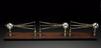
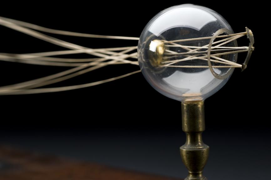
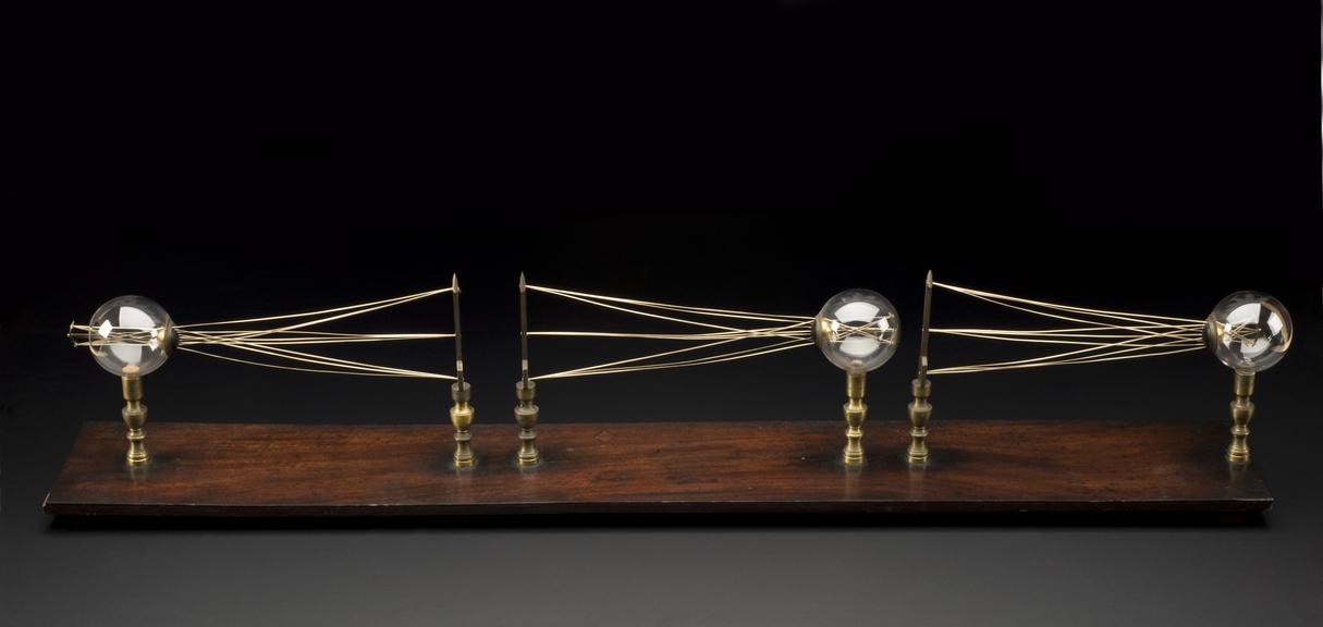
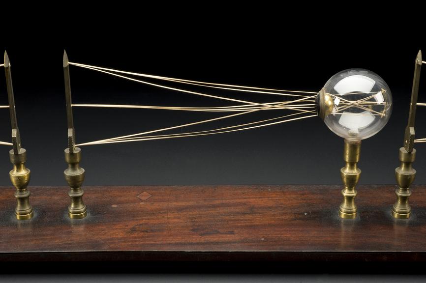
Eye defect teaching model, on wood stand, probably English, 1801-1900.
Showing how light enters the eye in long sight, normal sight and short sight; this is a 3-D model of the light diagrams seen in many science and medical textbooks. The model was probably used as a teaching aid for students studying the eye.
In normal sight, the light rays meet up at the retina, the sensitive part of the eye, and send a message to the brain through the optical nerve. Long sight (not being able to see close up) means that the light meets behind the retina; short sight (not being able to see far away) occurs when light meets up in front of the retina. Both conditions can be corrected with glasses. For a person with both long and short sight, bi-focal lenses can be used.
Details
- Category:
- Ophthalmology
- Collection:
- Sir Henry Wellcome's Museum Collection
- Object Number:
- A602310
- Measurements:
-
overall: 8.7402 x 6.6929 x 45.2756 in.; 222 x 170 x 1150 mm
- type:
- model
- credit:
- The Wellcome Trust
Related Objects
Rest spectacles

Pair of spectacles

Pair of spectacles
