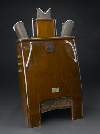


Polytome, universal tomograph, made by Philips Medical Systems in the 1950s.
Using this Polytome, a type of tomograph, a radiologist can X-ray parts of the body such as the skull or spine focussing on a particular bone joint. The patient is very precisely positioned and has to lie or sit completely still. The machine is very stable to ensure accurate exposures are obtained for a few seconds at a time. Parts of the tomograph then move around the patient to scan and film in a straight line, at different angles or in a preset pattern such as an ellipse to create a tomogram. Taking X-rays from different angles aid clinical diagnosis as the centre of the image is sharp but details outside the area of interest are blurred.
The Polytome was developed by Raymond Sans, and Jean Porcherat the Radiology department of the Assistance Publique hospitals in Paris, France. Their first experimental model was introduced in 1949. It incorporated the ideas of E M Bocage who proposed isolating the region of the body that needed to be X-rayed. Bocage had worked in a French military X-ray unit during the first world war. He patented his idea for the 'Technique and mechanism of a moving X-ray film' in 1921.
This machine was manufactured by Massiot Philips, based in Paris, France. This tomograph was used in Department of Radiology at the Radcliffe Infirmary, Oxford, England, from the late 1950s. Tomography was much used as a diagnostic tool until the late 1970s. It was replaced by the Computed Tomography (CT) scanner developed by Godfrey Hounsfield at EMI.
Details
- Category:
- Radiomedicine
- Object Number:
- 1998-15
- Materials:
- metal (unknown)
- Measurements:
-
overall (estimate): 2400 mm x 1850 mm x 1465 mm,
- type:
- tomograph
- credit:
- The Radcliffe Infirmary
