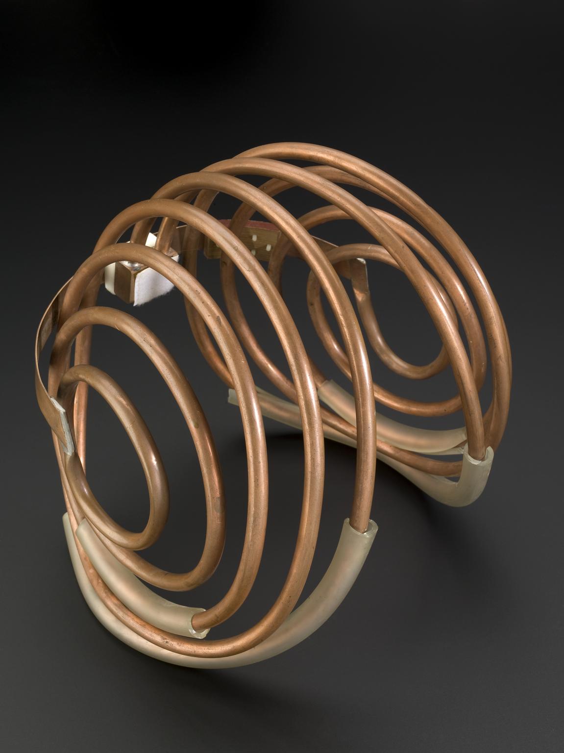
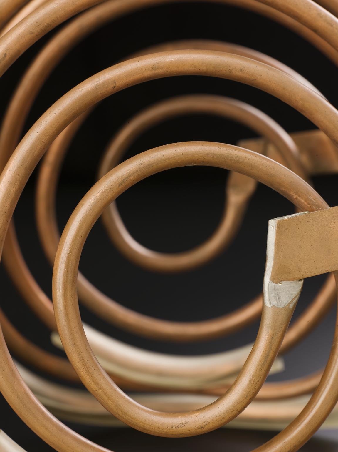
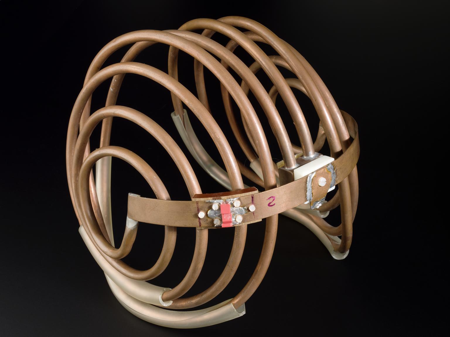
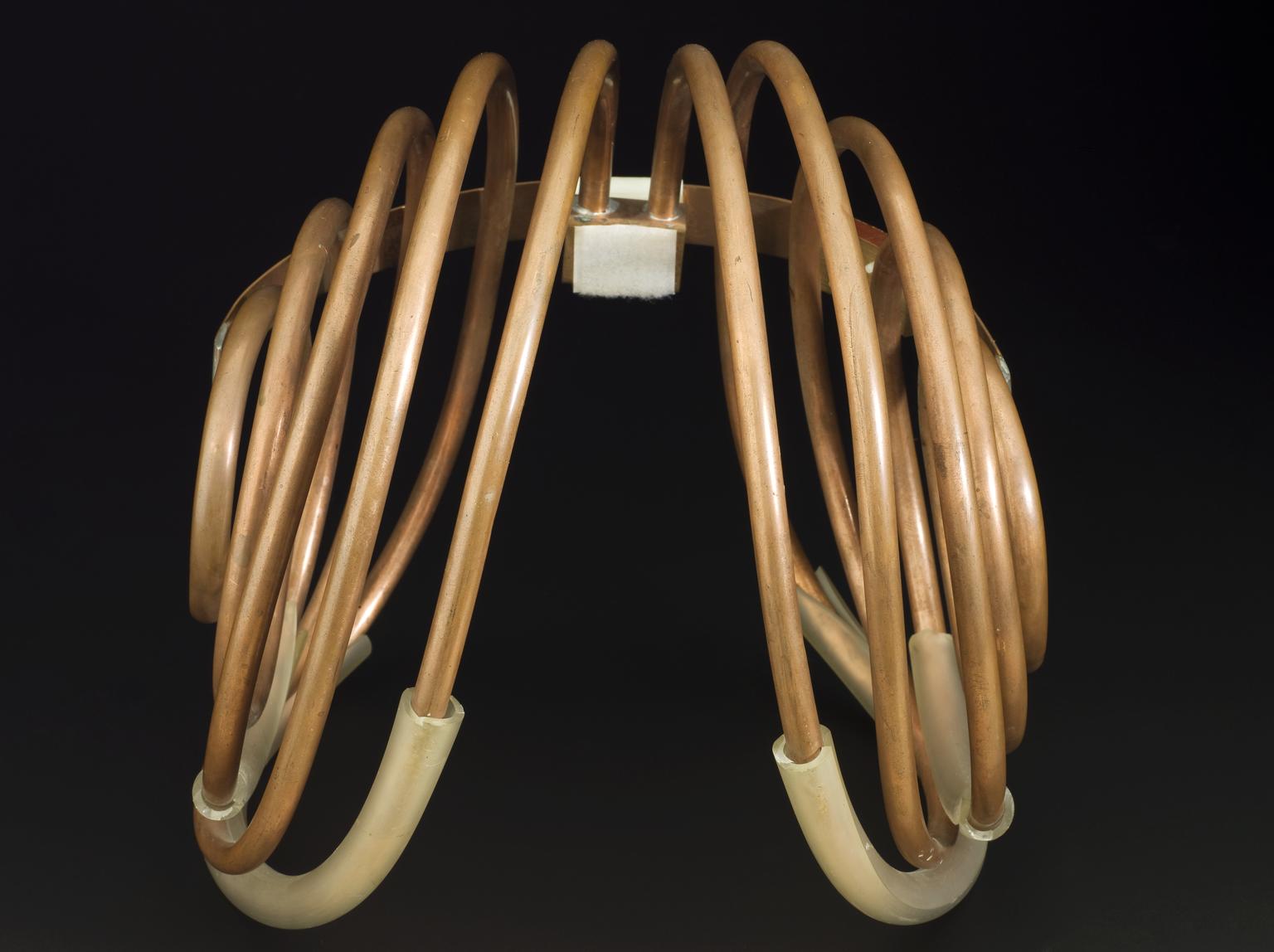
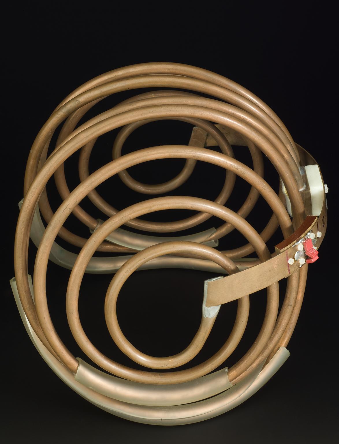
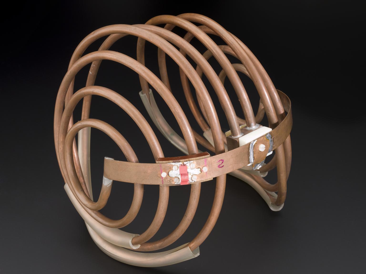
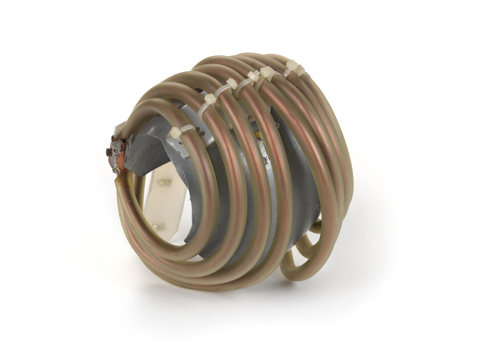
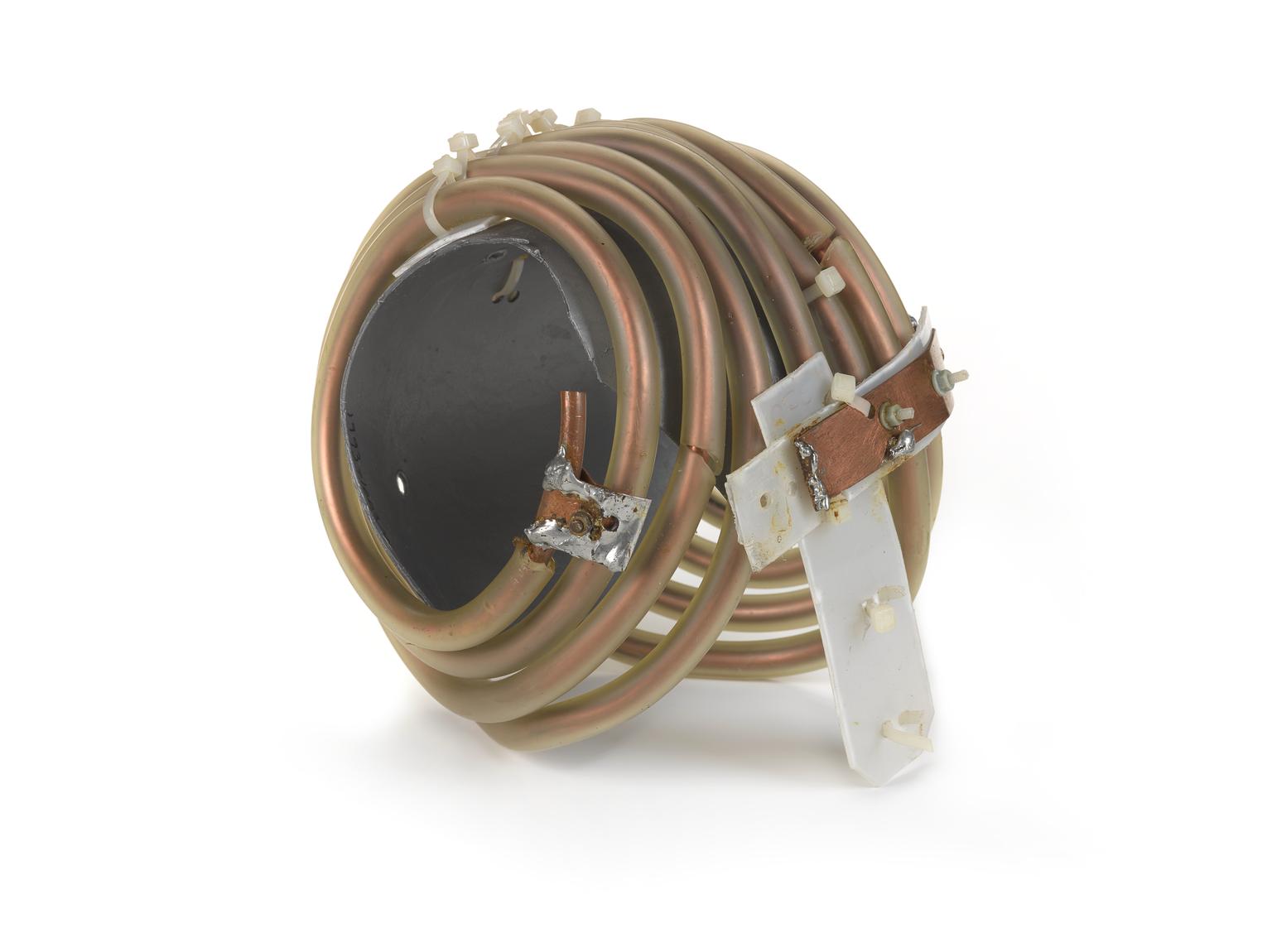
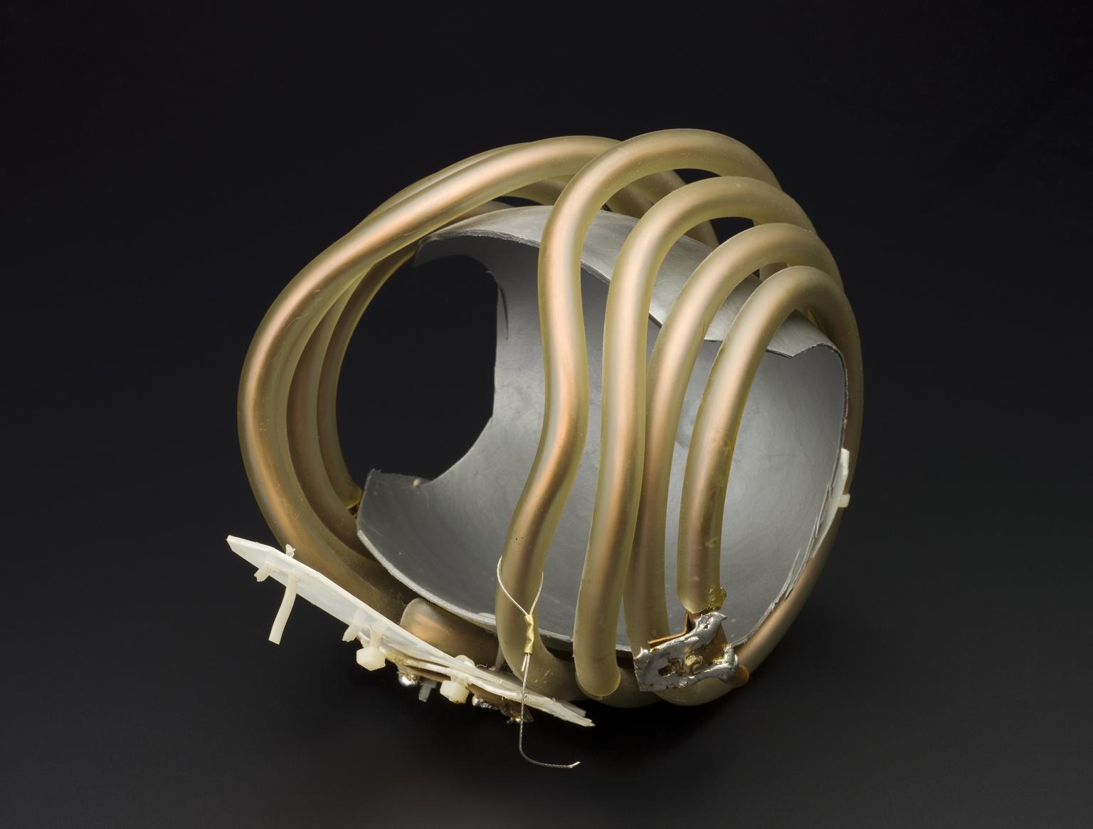
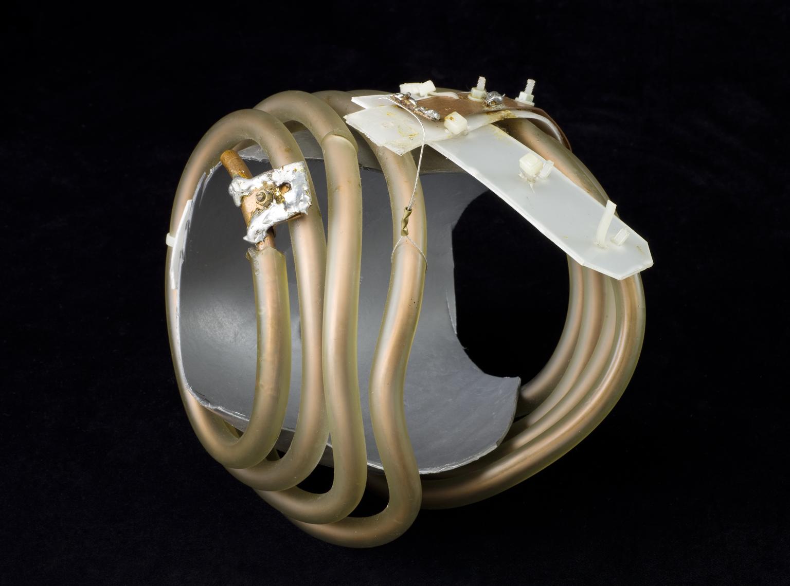
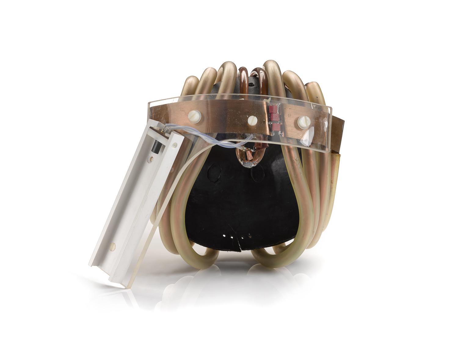
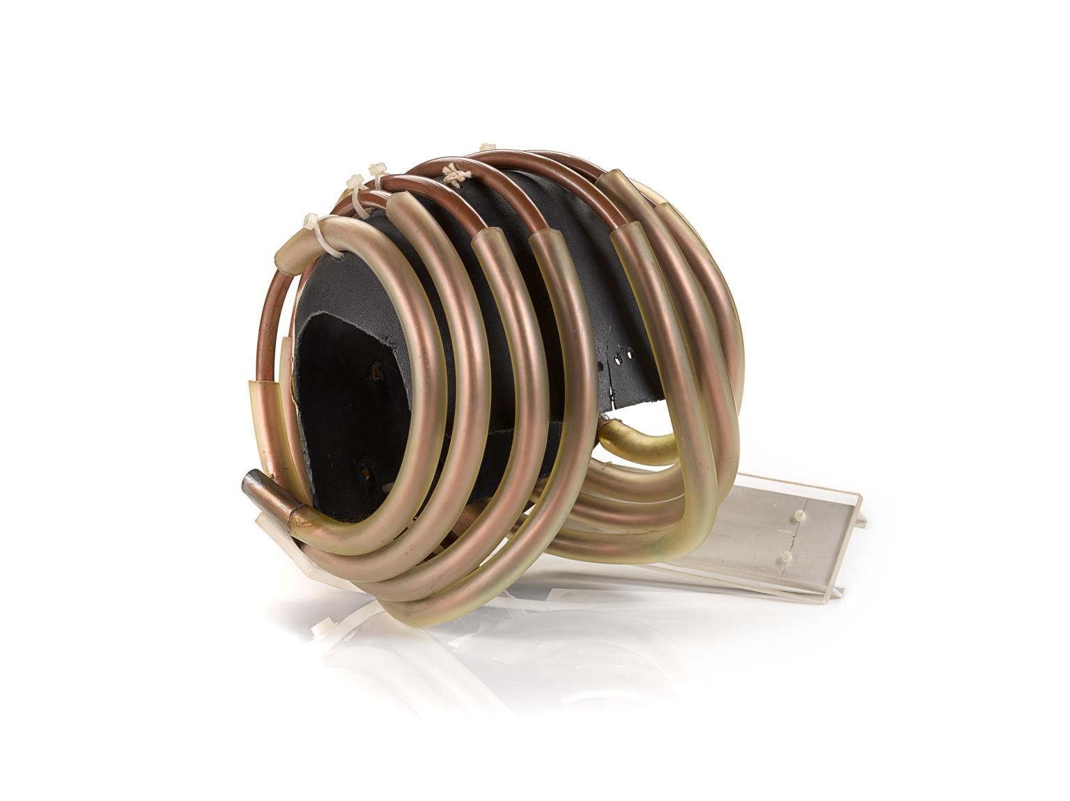
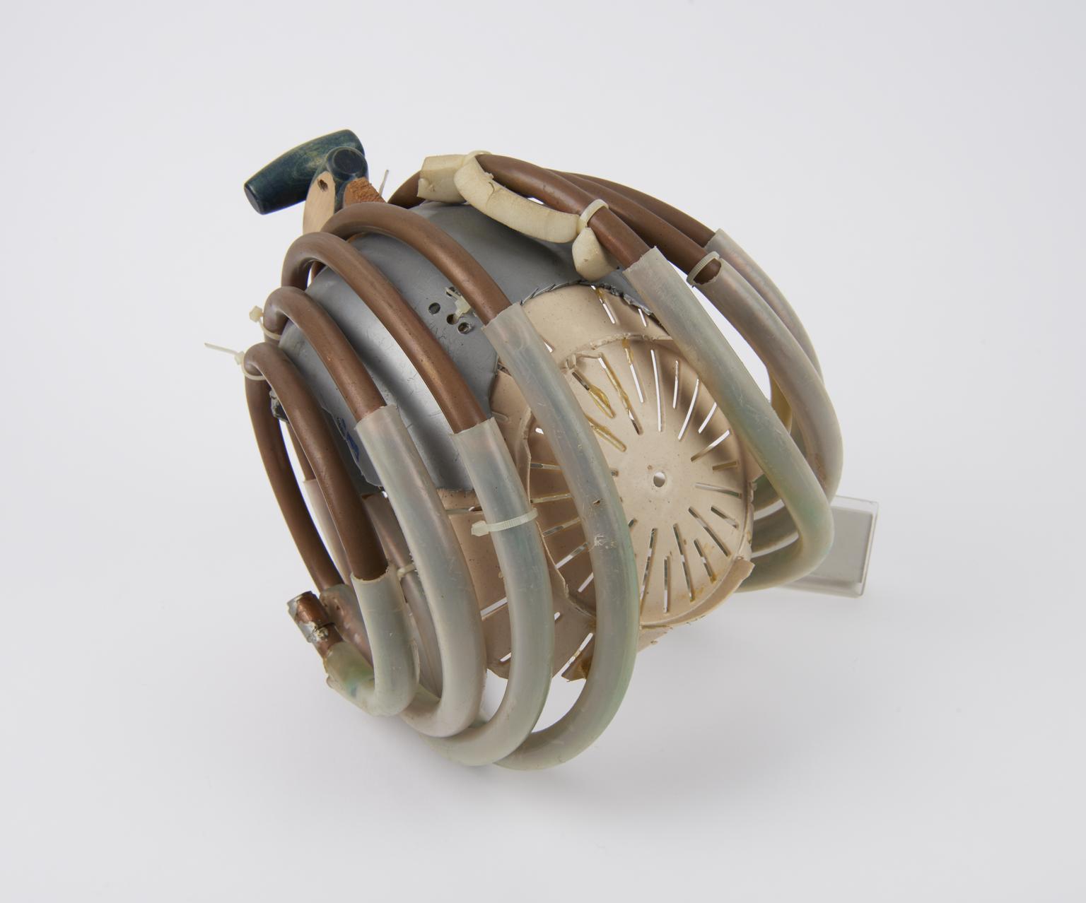
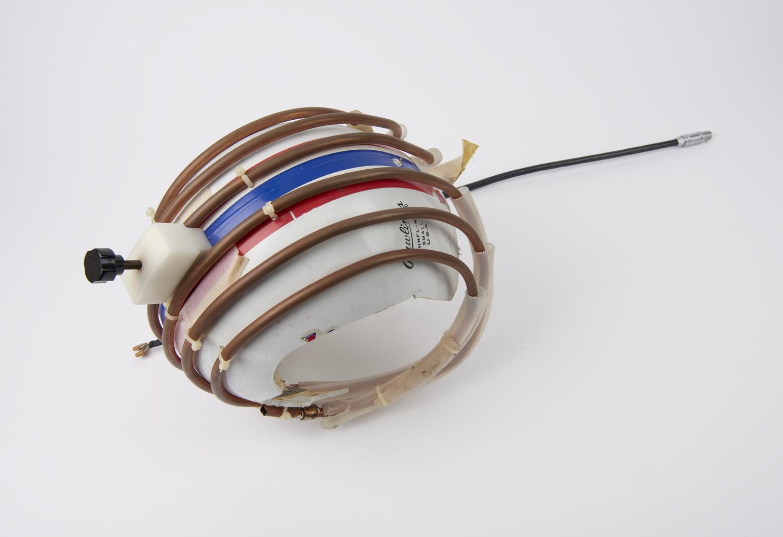
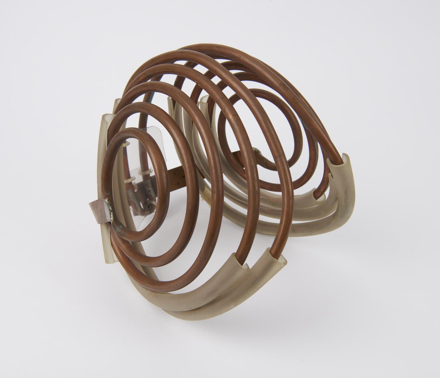
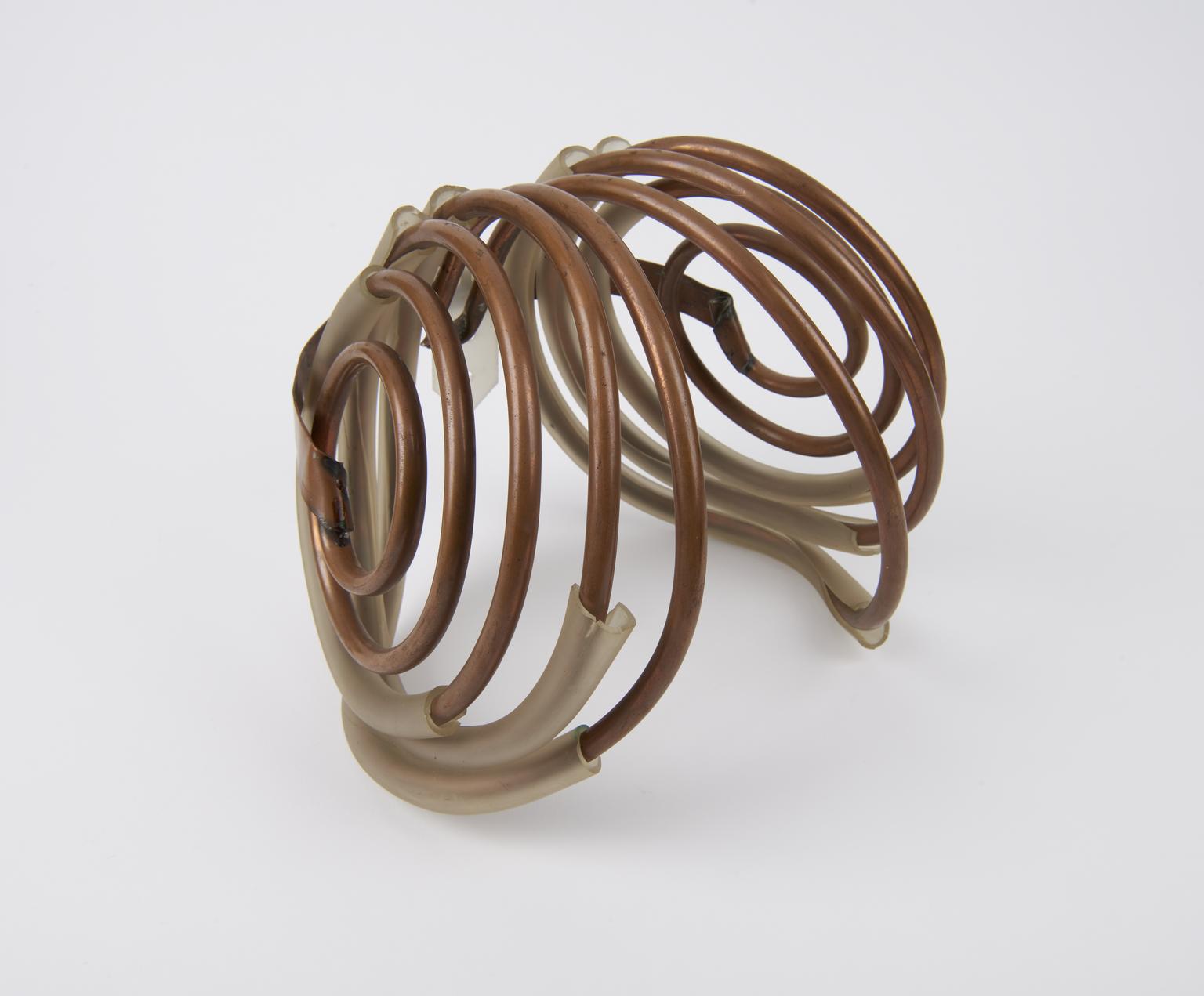
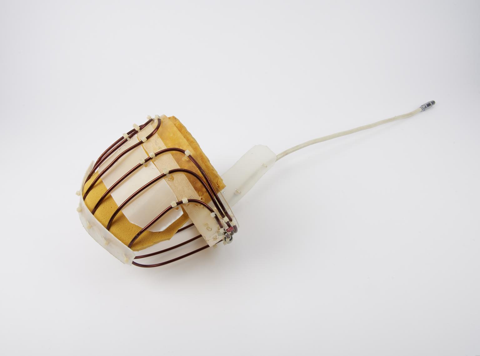
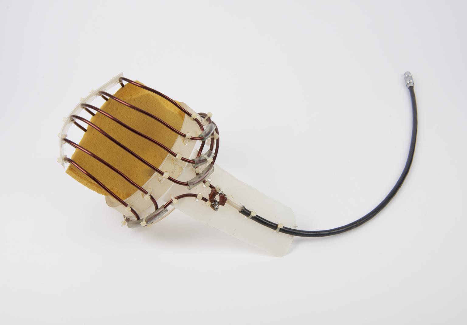
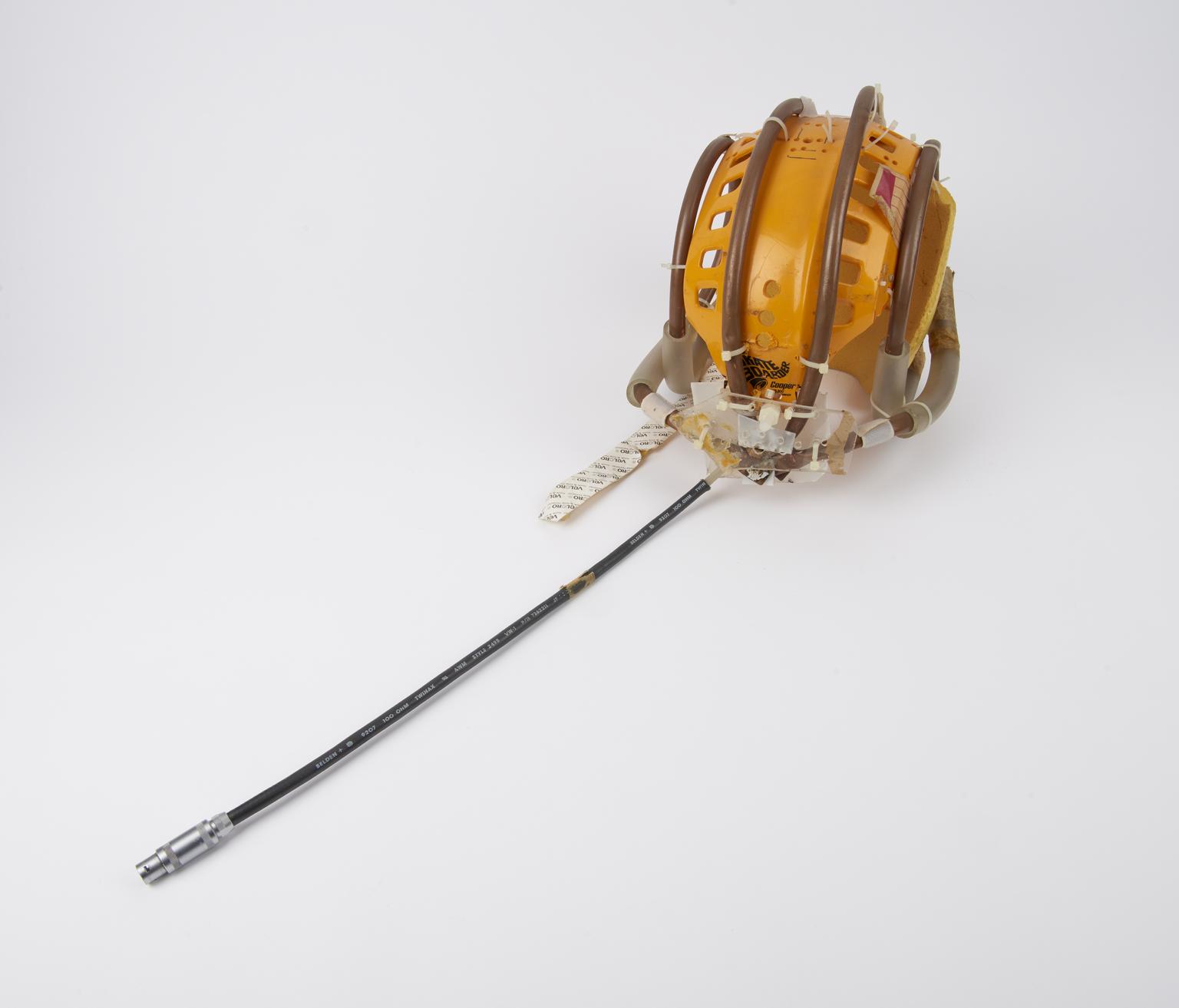
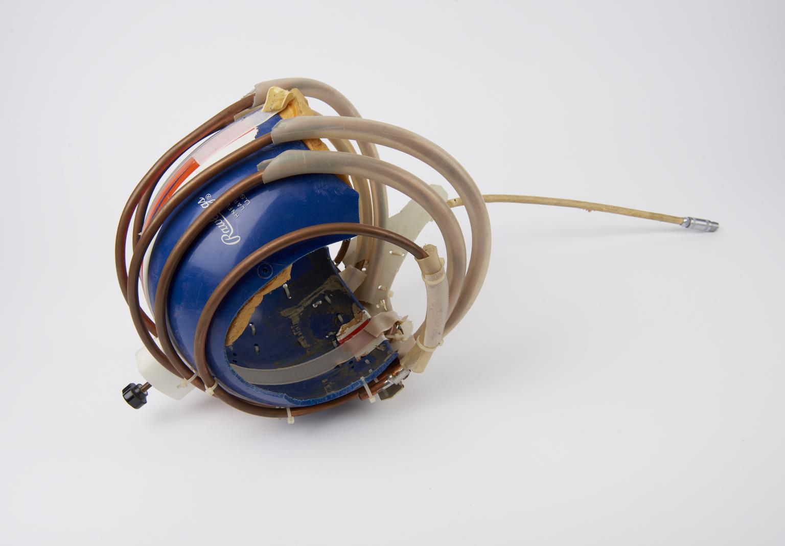
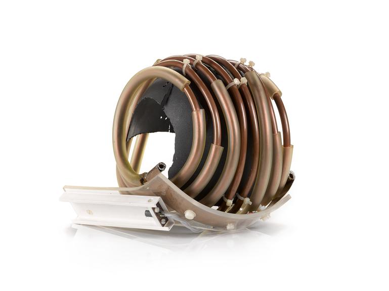
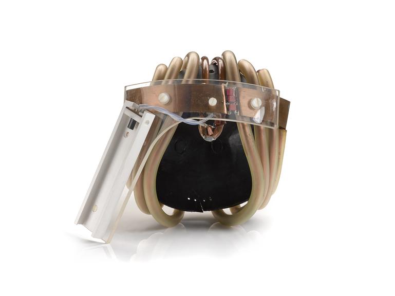
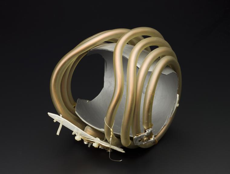
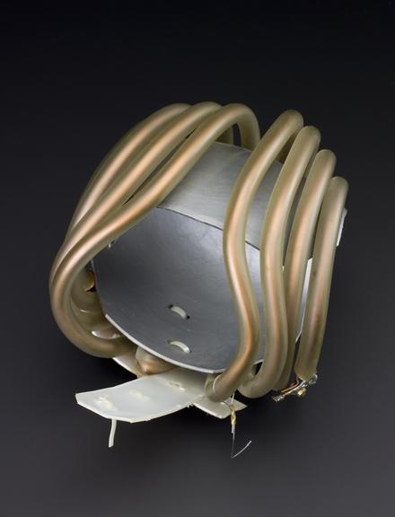
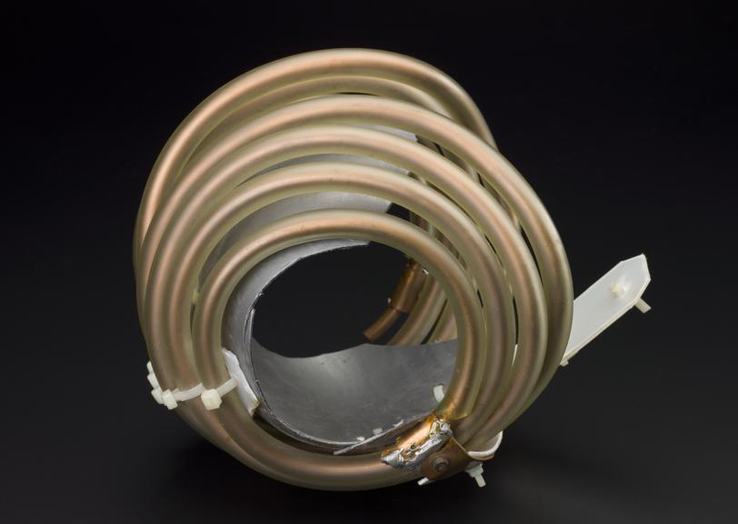
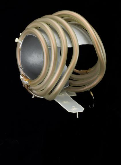
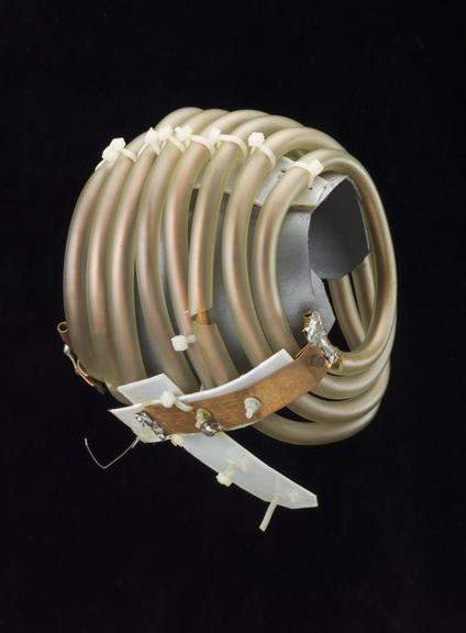
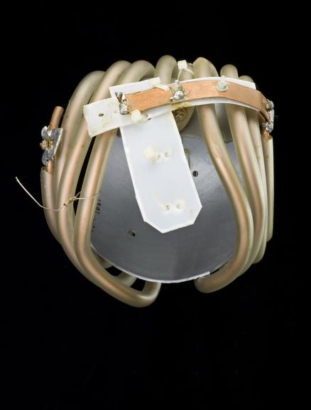
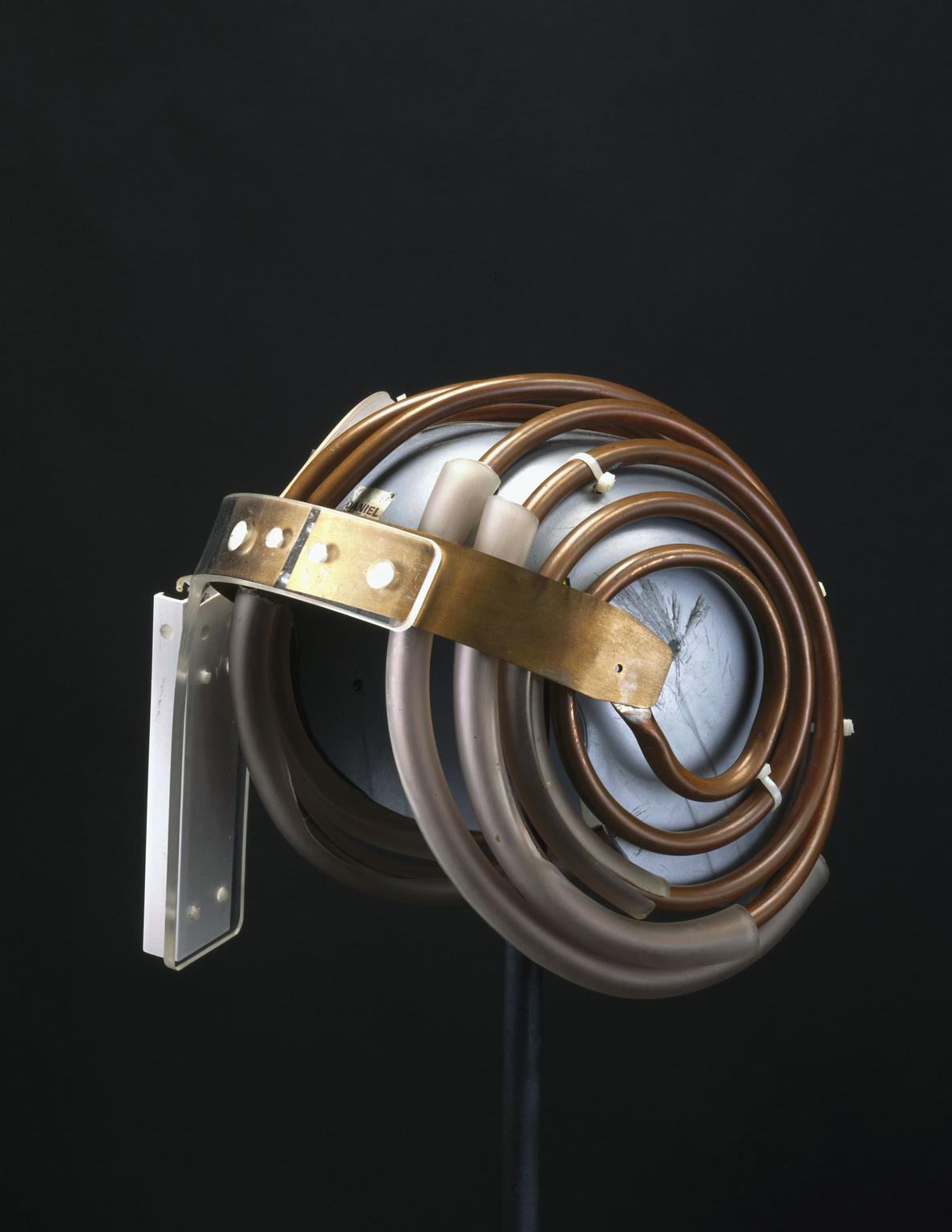
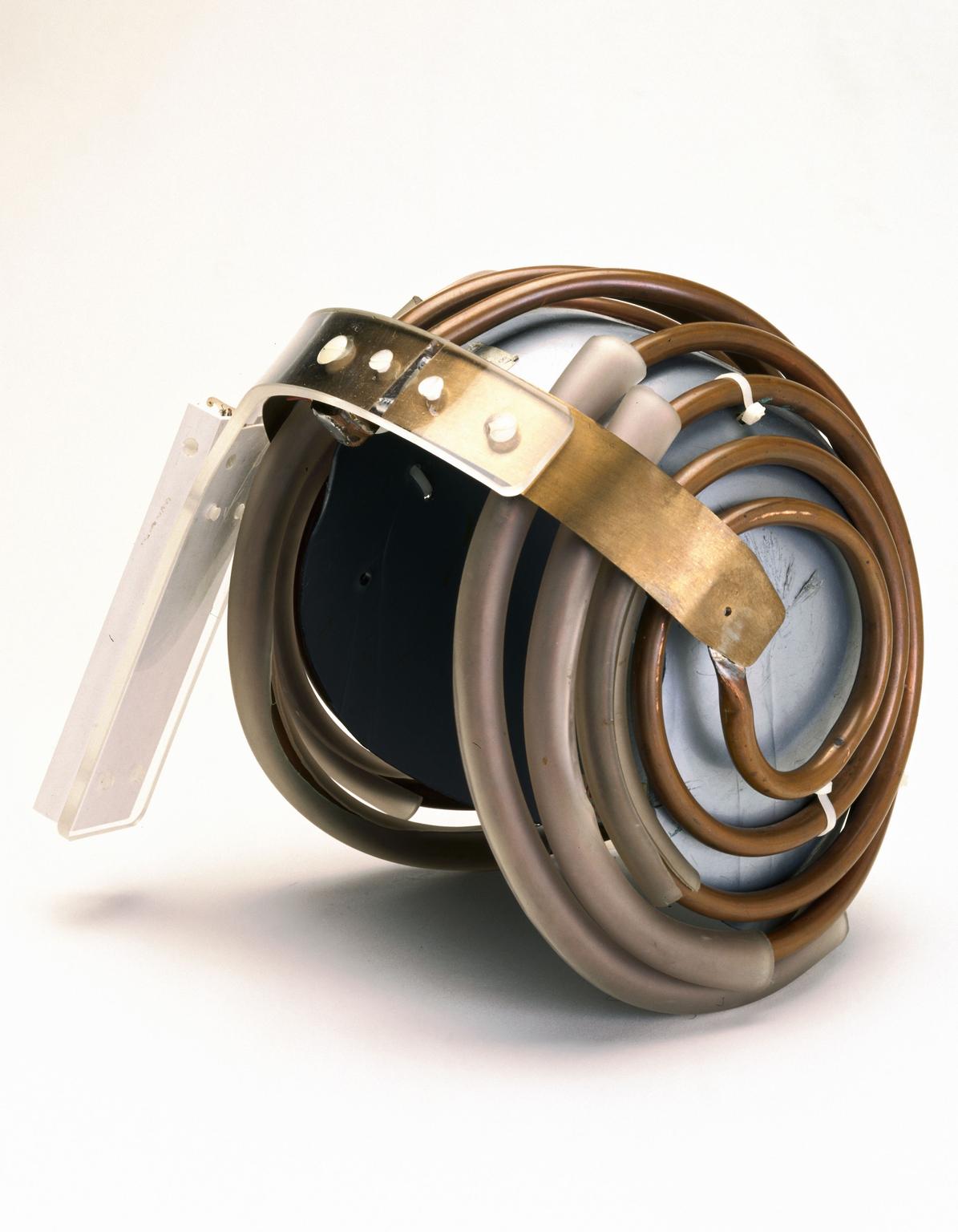
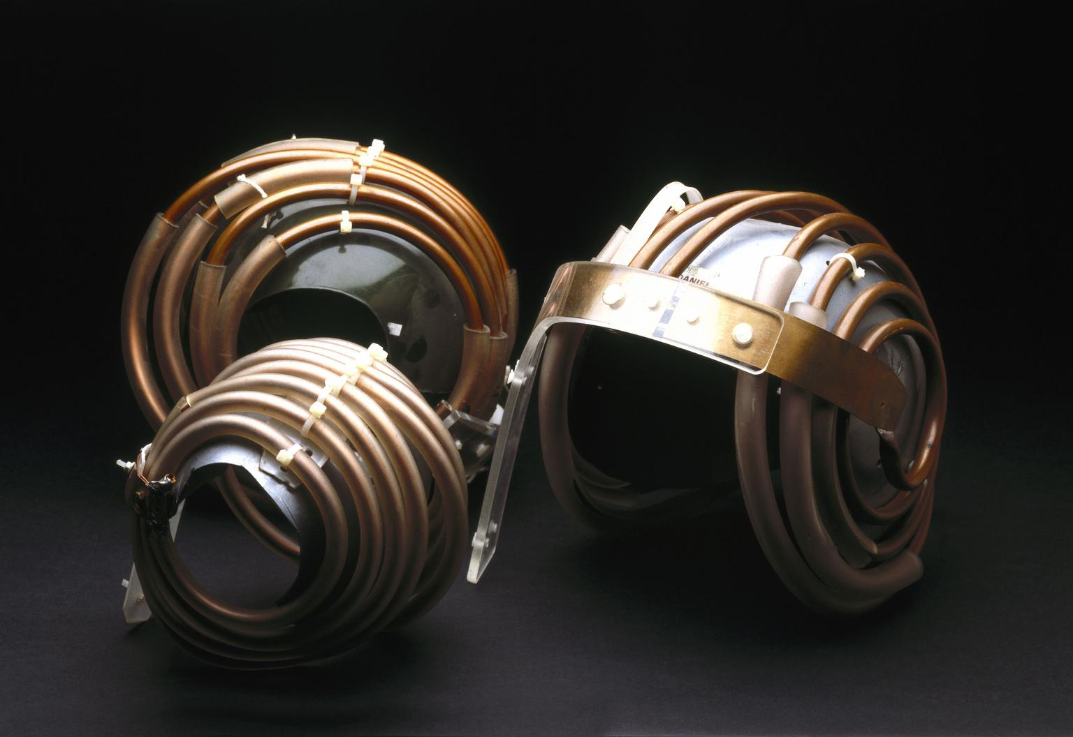
'Jedi' helmet, used with Cryogenic Magnetic Resonance Imaging Machine

Made:
1980 in United States
maker:
Data General
and
Data General
and
Group of 'Jedi' helmets for use in MRI scanning of the brain, 1984. General view. Black background. One of a group of 'Jedi' helmets for use in MRI scanning of the brain, 1984. Front three quarter view. White background. One of a group of 'Jedi' helmets for use in MRI scanning of the brain, 1984. Front three quarter view. Black background.

Buy this image as a print 

Buy

License this image for commercial use at Science and Society Picture Library

License

Buy this image as a print 

Buy

License this image for commercial use at Science and Society Picture Library

License

Buy this image as a print 

Buy

License this image for commercial use at Science and Society Picture Library

License

Group of 'Jedi' helmets for use in MRI scanning of the brain, 1984. General view. Black background.
Science Museum Group Collection
© The Board of Trustees of the Science Museum

One of a group of 'Jedi' helmets for use in MRI scanning of the brain, 1984. Front three quarter view. White background.
Science Museum Group Collection
© The Board of Trustees of the Science Museum

One of a group of 'Jedi' helmets for use in MRI scanning of the brain, 1984. Front three quarter view. Black background.
Science Museum Group Collection
© The Board of Trustees of the Science Museum

One of five 'Jedi helmets,' silver inner helmet, used with Cryogenic Magnetic Resonance Imaging Machine and associated computing equipment by Data General, United States,1980.

To take MRI (Magnetic Resonance Imaging) scans of the brain, these helmets were worn by children and adults. The coils are aerials for picking up MRI signals. Naming them after the Jedi knights in Star Wars films encouraged children to put them on and not to be frightened.

MRI builds up a picture of the human body by using high frequency radio waves known as NMR (Nuclear Magnetic Resonance). MRI does not expose the body to radiation or invasive surgery and it can image soft tissues more effectively than X-ray-based methods.

Details

Category:
Radiomedicine
Object Number:
1993-1003/2 Pt1
Materials:
brass (copper, zinc alloy), copper (alloy), electrical components and plastic (unidentified)
Measurements:
overall: 320 mm x 250 mm x 250 mm, 4.595 kg
type:
component - object
taxonomy:
  • furnishing and equipment
  • tools & equipment
credit:
Hammersmith Hospital