


![]() This image is released under a CC BY-NC-SA 4.0 Licence
This image is released under a CC BY-NC-SA 4.0 Licence
Buy this image as a print
BuyLicense this image for commercial use at Science and Society Picture Library
License![]() This image is released under a CC BY-NC-SA 4.0 Licence
This image is released under a CC BY-NC-SA 4.0 Licence
Buy this image as a print
BuyLicense this image for commercial use at Science and Society Picture Library
License![]() This image is released under a CC BY-NC-SA 4.0 Licence
This image is released under a CC BY-NC-SA 4.0 Licence
Buy this image as a print
BuyLicense this image for commercial use at Science and Society Picture Library
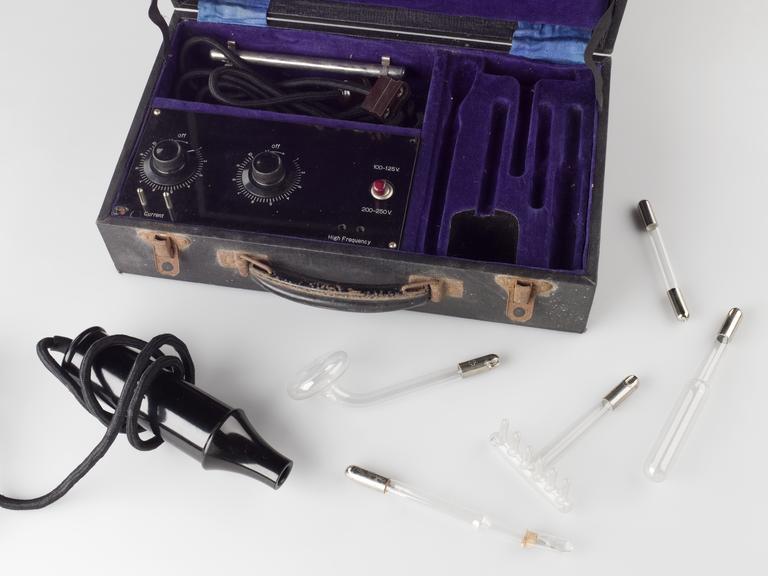
LicenseHigh frequency (violet ray) apparatus as used by Chisholm
Science Museum Group
© The Board of Trustees of the Science Museum
High frequency (violet ray) apparatus as used by Chisholm
Science Museum Group Collection
© The Board of Trustees of the Science Museum
High frequency (violet ray) apparatus as used by Chisholm
Science Museum Group Collection
© The Board of Trustees of the Science Museum
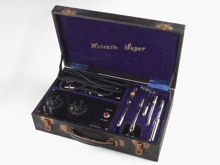
High frequency (violet ray) apparatus as used by Chisholm Williams, with five electrode accessories, c.1880-1928
Powered by mains electricity supply, this set was used to apply high frequency electrical sparks to the body using the glass vacuum electrodes. Known as violet ray treatment, the purple, red, or blue electric sparks were believed to relieve aches and pains in the muscles, internal organs, and the nerves (rheumatism) by massaging the surface of the skin with the electrode.
This kit was owned by Chisholm Williams (1866-1928) was an early adopter of X-ray and electrotherapy. Within weeks of hearing about the discovery of X-rays in 1896, he managed to take an X-ray of his fingers. he was in charge of the X-ray department of West London Hospital and lectured in radiology. He served in the Royal Army Medical Corps as captain and electrotherapeutist. Due to the prolonged exposure to X-rays, when their dangers were unknown, led to Williams’ hand being damaged and two fingers on his other hand amputated.
Details
- Category:
- Therapeutics
- Object Number:
- 2008-36
- Materials:
- wood, glass and electronic components
- Measurements:
-
overall: 93 mm x 350 mm x 240 mm, 2.24 kg
overall (open): 277 mm x 350 mm x 270 mm, 2.24 kg
- credit:
- Gift of Mr Christopher Chisolm Barry