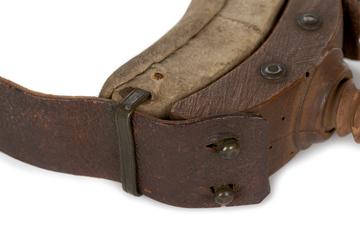
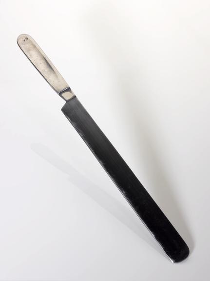
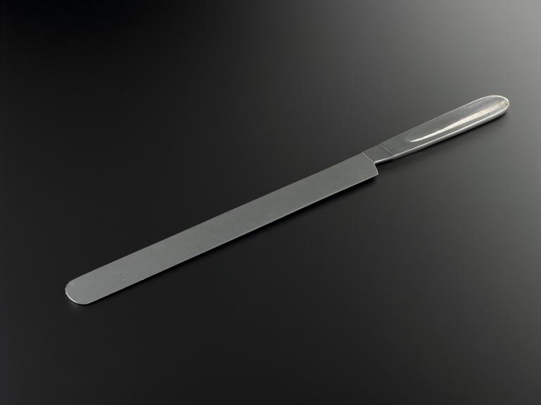
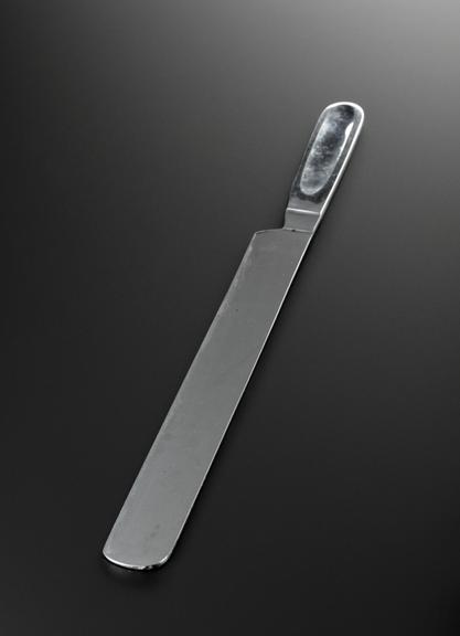
Pathologists' brain knife
- Made:
- circa 2016 in unknown
- maker:
- Unknown


![]() This image is released under a CC BY-NC-SA 4.0
Licence
This image is released under a CC BY-NC-SA 4.0
Licence
License this image for commercial use at Science and Society Picture Library
License![]() This image is released under a CC BY-NC-SA 4.0
Licence
This image is released under a CC BY-NC-SA 4.0
Licence
License this image for commercial use at Science and Society Picture Library
License![]() This image is released under a CC BY-NC-SA 4.0
Licence
This image is released under a CC BY-NC-SA 4.0
Licence
License this image for commercial use at Science and Society Picture Library
License
Science Museum Group Collection
© The Board of Trustees of the Science Museum
Brain knife used by pathologists to slice thin sections of the
Science Museum Group Collection
© The Board of Trustees of the Science Museum
Brain knife used by pathologists to slice thin sections of the
Science Museum Group Collection
© The Board of Trustees of the Science Museum
Brain knife used by pathologists to slice thin sections of the brain during post mortem, collected from the Department of Cellular Pathology at the John Radcliffe Hospital, 2016.
Post-mortems are completed to establish cause of death on around 20 percent of people who die in Britain each year. After careful external inspection of the body, specialised tools are used to open it up to locate signs of disease or injury.
Equipment also protects the pathologist while they are working. While many of the tools and practices of post mortem have remained unchanged for years, digital imaging technologies are gradually being used to complete post-mortem examinations.
Details
- Category:
- Surgery
- Object Number:
- 2016-472
- Materials:
- stainless steel
- type:
- brain knife
- credit:
- Gift of John Radcliffe Hospital