![]() This image is released under a CC BY-NC-SA 4.0
Licence
This image is released under a CC BY-NC-SA 4.0
Licence
License this image for commercial use at Science and Society Picture Library
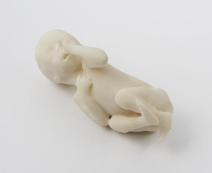
License3D printed model of a normal fetus at 22 weeks of gestation
Science Museum Group
© The Board of Trustees of the Science Museum
3D printed model of a healthy fetus at 22 weeks of gestation made by the Centre for Advanced Additive Manufacturing (AdAM) at the University of Sheffield.
Made by researchers at the University of Sheffield, this model shows one way 3D printing is being used in medicine. Part of a teaching set for radiologists, these true to size models are used to help improve their understanding of fetal anatomy and their skills in reading in-utero MRI (magnetic resonance imaging) scans. MRI scans produce layers and layers of 2D images which have been combined to create and print this 3D model. By turning these images into something physical, the researchers hope to help radiologists visualise and understand what they see in scans.
Details
- Category:
- Radiomedicine
- Object Number:
- 2019-467
- Materials:
- thermoplastic polymer (Nylon 12)
- Measurements:
-
overall: 200 mm x 110 mm x 80 mm,
- type:
- model