![]() This image is released under a CC BY-NC-SA 4.0
Licence
This image is released under a CC BY-NC-SA 4.0
Licence
Buy this image as a print
BuyLicense this image for commercial use at Science and Society Picture Library
License3D printed model of an abnormal fetal brain showing
Science Museum Group
© The Board of Trustees of the Science Museum
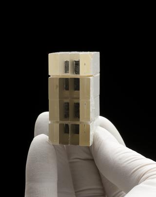
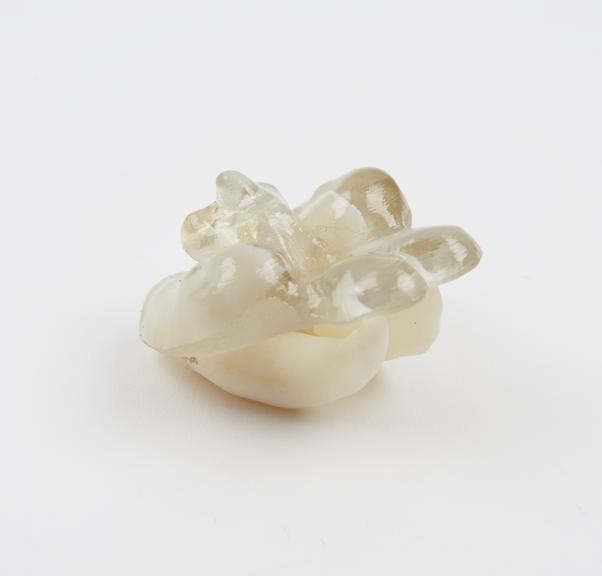
3D printed model of a fetal brain showing ventriculomegaly, the clear resin shows the structure of the brain, while the white resin shows the enlarged ventricular system. 21 weeks of gestation, made by the Centre for Advanced Additive Manufacturing (AdAM) at the University of Sheffield, 2012-2018
Made by researchers at the University of Sheffield, this model shows one way 3D printing is being used in medicine. Part of a teaching set for radiologists, these true to size models are used to help improve their understanding of fetal anatomy and in-utero MRI (magnetic resonance imaging) scanning. MRI scans produce layers and layers of 2D images which have been combined to create and print this 3D model. By turning these images into something physical, the researchers hope to help radiologists visualise and understand what they see in scans. This model even uses two different materials to differentiate between the structure of the brain and the ventricles. Within the set, there are examples of brain malformations at several gestational ages which can be compared to examples showing healthy brain development.
Ventriculomegaly is a condition where the ventricles of the brain, which produce and contain cerebral spinal fluid are enlarged. The symptoms vary hugely and depend on how affected the brain is. It can be diagnosed during routine ultrasounds.
Details
- Category:
- Radiomedicine
- Object Number:
- 2019-478
- Measurements:
-
overall: 65 mm x 55 mm x 50 mm,
- type:
- model