![]() This image is released under a CC BY-NC-SA 4.0
Licence
This image is released under a CC BY-NC-SA 4.0
Licence
Buy this image as a print
BuyLicense this image for commercial use at Science and Society Picture Library
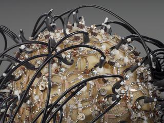
License3D printed model of a normal fetal brain in three sections and
Science Museum Group
© The Board of Trustees of the Science Museum
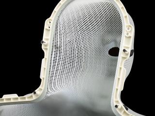
3D printed model of a healthy fetal brain in three sections and overlaid with MRI images at 21 weeks gestation, made by the Centre for Advanced Additive Manufacturing (AdAM) at the University of Sheffield, 2012-2018
Made by researchers at the University of Sheffield, this model shows one way 3D printing is being used in medicine. Part of a teaching set for radiologists, these true to size models are used to help improve their understanding of fetal anatomy and in-utero MRI (magnetic resonance imaging) scanning. MRI scans produce layers and layers of 2D images which have been combined to create and print this 3D model. By turning these images into something physical, the researchers hope to help radiologists visualise and understand what they see in scans. Within the set, there are examples of brain malformations at several gestational ages which can be compared to examples showing healthy brain development.
This model shows how the 3D printing can also be combined with the original MRI scans to help radiologists understand the relationship between the model and the internal structure of the brain.
Details
- Category:
- Radiomedicine
- Object Number:
- 2019-479
- Measurements:
-
overall: 75 mm x 55 mm x 60 mm,
- type:
- model