Case for teaching set of 3D printed models of fetal brains
- Made:
- Unknown in unknown place
![]() This image is released under a CC BY-NC-SA 4.0 Licence
This image is released under a CC BY-NC-SA 4.0 Licence
Buy this image as a print
BuyLicense this image for commercial use at Science and Society Picture Library
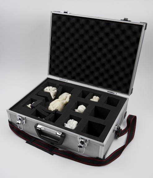
LicenseCase to transport teaching set of 3D printed models of fetal
Science Museum Group
© The Board of Trustees of the Science Museum
Case to transport teaching set of 3D printed models of fetal brains, with paper manual titled 'An introduction to 3D MR imaging of the fetal brain', made by the Centre for Advanced Additive Manufacturing (AdAM) at the University of Sheffield, 2012-2018
Made by researchers at the University of Sheffield, these models show one way 3D printing is being used in medicine. Part of a teaching set for radiologists, these true to size models are used to help improve their understanding of fetal anatomy and in-utero MRI (magnetic resonance imaging) scanning. MRI scans produce layers and layers of 2D images which have been combined to create and print this 3D model. By turning these images into something physical, the researchers hope to help radiologists visualise and understand what they see in scans. Within the set, there are examples of brain malformations at several gestational ages which can be compared to examples showing healthy brain development. The set is accompanied by a leaflet containing sample reports, images and background information about the conditions included.
Details
- Category:
- Radiomedicine
- Object Number:
- 2019-480
- Materials:
- metal (unknown), textile and plastic (unidentified)
- Measurements:
-
overall (closed): 160 mm x 455 mm x 350 mm,
- type:
- case