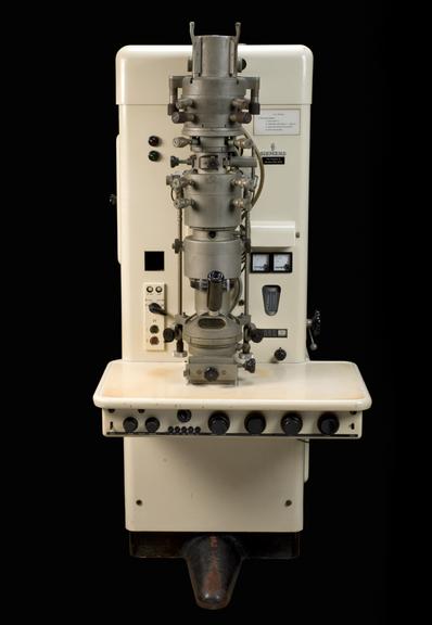
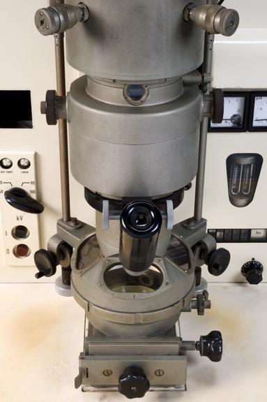
Siemens Elmiskop 1 electron microscope, the first electron microscope used in the Department of Human Anatomy at Oxford, c. 1950
An electron microscope uses a beam of electrons, instead of light, to produce highly magnified images of objects. Electrons have a much smaller wavelength than visible light. This allows a much higher resolution to be achieved. The instrument was first developed by Ernst Ruska (1906-88) in Berlin in the early 1930s. The Siemens company made the first commercial machines later in the decade. Ruska returned to designing electron microscopes with Siemens after the Second World War. The Elmiskop 1 of 1954 was their first innovation. It became a worldwide success. It was the first microscope with a ‘double condenser’. This allowed routine electron diffraction. This microscope was the first electron microscope used in the Department of Human Anatomy at Oxford University.