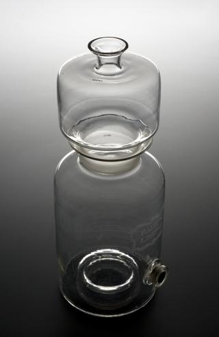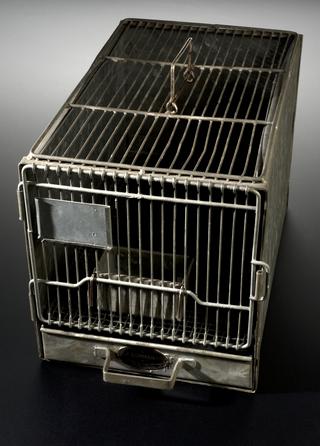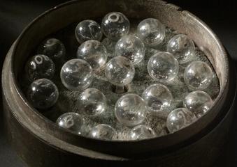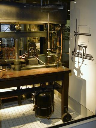
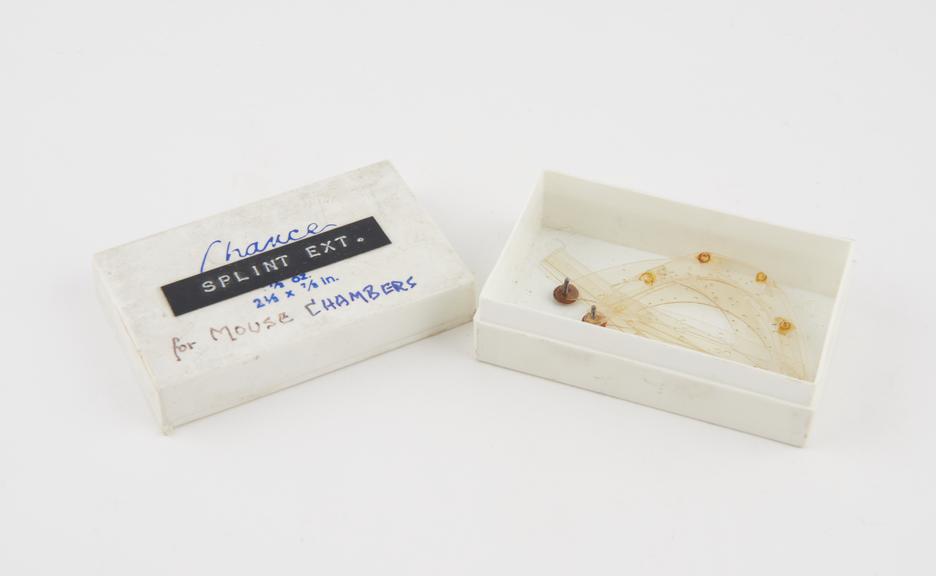
Perspex models in plastic box labelled 'Splint Ext.', parts of mouse chambers used for the study of circulation in animals by Dr. Glenn Algire at the National Cancer Institute, 1940-1950
During a 17 year career at the National Cancer Institute in the United States, Glenn Algire (1907-1950) studied cells and tumours in living animals. His fascination with cells and tissues began during his internship after graduating from medical school. During a 2 year fellowship, he developed a way of inserting transparent chambers into the skin of anaesthetised mice. While others had attempted to do so in rabbits, Algire was one the first to do so in mice. His techniques allowed him to develop methods of studying pressure in blood vessels, and ways of observing individual cells including using time-lapse photography. Tumours in mice can mimic human cancers so are often used in research – although under strict rules and supervision.
These examples belonged to Peter Alexander George Monro (1919-2005), an anatomist, lecturer, and Fellow of St John's College, University of Cambridge. He invented and devised innovative methods of visualising blood flow in vivo (living) animals and found ways of measuring the velocity of red blood cells. Monro’s work looked mainly at rabbit ear chambers rather than mice but it is likely that he learnt from the work, success and failures of others, including Algire.
