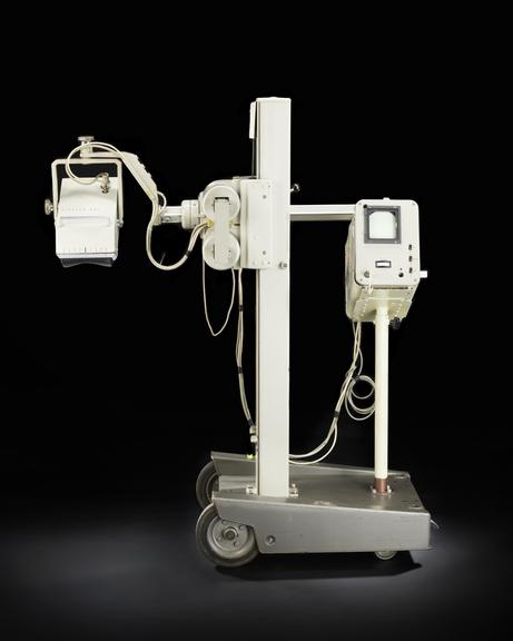
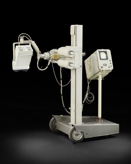
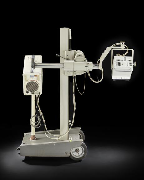
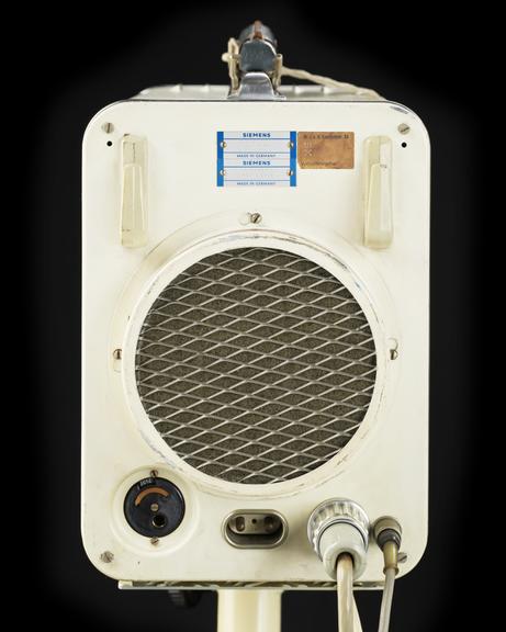
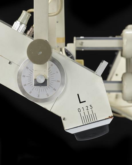
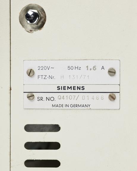
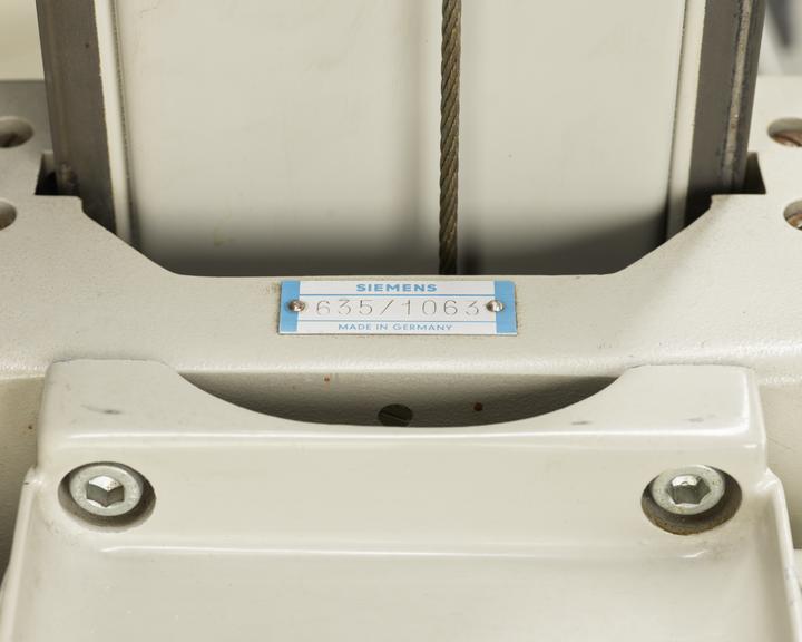
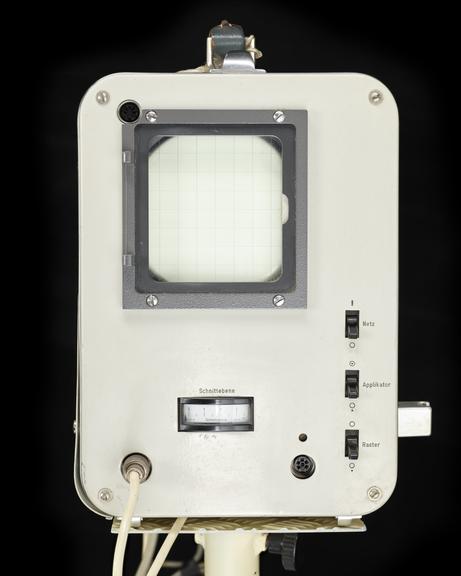
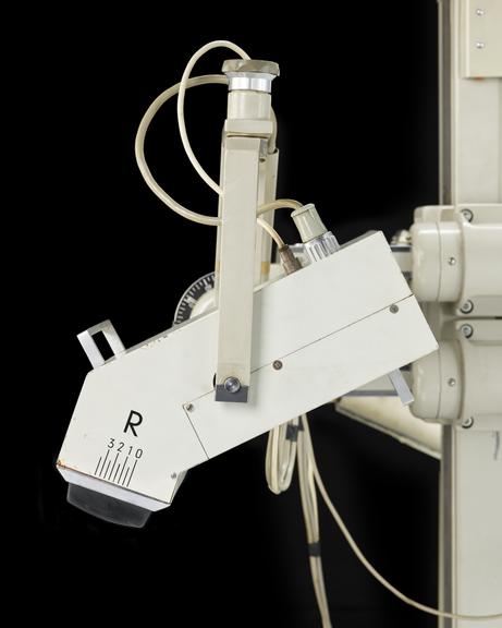
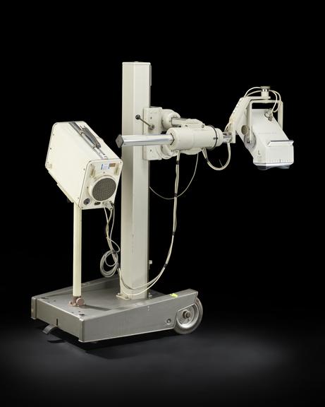
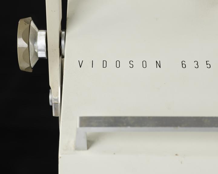
Mechanical real-time scanner ‘Vidoson 635’

Made:
1965 in Germany
maker:
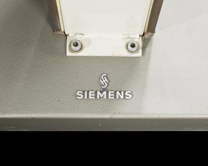
Siemens AG

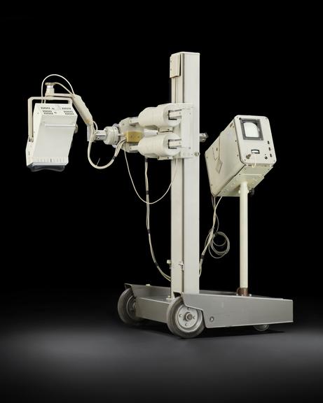
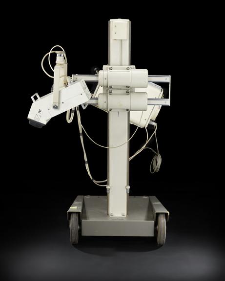
Mechanical real-time scanner ‘Vidoson 635’, Siemens, Germany, 1965

Developed by Siemens engineers, including Richard Soldner (b1935), the ‘Vidoson 635’ was the first ultrasound machine to show real-time observation inside of people’s bodies. 2D grey scale images showed movement and anatomical structures. Soldner was trying to solve the issue of improved resolution, showing different grey scales and faster scanning in breast screening. The Vidoson was his solution.

After unsuccessful initial testing in 1962 and further setbacks in 1965, a team including Hans J Hollander at the gynaecological hospital in Münster looking for ways to perform abdominal ultrasound diagnostics. Prototypes of the Vidoscan were sent to the hospital with Richard Soldner and engineer Walter Krause. Beginning with reproducing the work of those working with ultrasound to diagnose gynaecological conditions, the team extended its use to foetal and obstetric imagining. For the first time, foetal heartbeats and breathing could be seen in real time. Prior to the use of ultrasound, X-rays were used and only showed static images. Medical opinion of up the 1950s were that low doses of x-rays were safe. Alice Stewart’s work demonstrated the link between foetal x-rays and childhood cancers. Despite publishing her study in 1958, it took years for her work to be taken seriously.

This success in obstetric and foetal imagining led to the Vidoson being put into production. Around 3000 units, including this one were sold and used across Europe. By 1980, the Vidoson was no longer made, being overtaken by improved technologies.

Details

Category:
Clinical Diagnosis
Object Number:
2023-469
Materials:
metal, electronic components, plastic, rubber and lead
Measurements:
overall: 1650 mm x 1830 mm x 1100 mm, 200 kg
type:
ultrasound scanner

Parts

Mechanical real-time scanner ‘Vidoson 635’

Mechanical real-time scanner ‘Vidoson 635’

Mechanical real-time scanner ‘Vidoson 635’, Siemens, Germany 1965

More

Developed by Siemens engineers, including Richard Soldner (b1935), the ‘Vidoson 635’ was the first ultrasound machine to show real-time observation inside of people’s bodies. 2D grey scale images showed movement and anatomical structures. Soldner was trying to solve the issue of improved resolution, showing different grey scales and faster scanning in breast screening. The Vidoson was his solution.

After unsuccessful initial testing in 1962 and further setbacks in 1965, a team including Hans J Hollander at the gynaecological hospital in Münster looking for ways to perform abdominal ultrasound diagnostics. Prototypes of the Vidoscan were sent to the hospital with Richard Soldner and engineer Walter Krause. Beginning with reproducing the work of those working with ultrasound to diagnose gynaecological conditions, the team extended its use to foetal and obstetric imagining. For the first time, foetal heartbeats and breathing could be seen in real time. Prior to the use of ultrasound, X-rays were used and only showed static images. Medical opinion of up the 1950s were that low doses of x-rays were safe. Alice Stewart’s work demonstrated the link between foetal x-rays and childhood cancers. Despite publishing her study in 1958, it took years for her work to be taken seriously.

This success in obstetric and foetal imagining led to the Vidoson being put into production. Around 3000 units, including this one were sold and used across Europe. By 1980, the Vidoson was no longer made, being overtaken by improved technologies.

Measurements:
overall: 1650 mm x 1830 mm x 1100 mm, 200 kg
Materials:
metal , electronic components , plastic , rubber and lead
Object Number:
2023-469/1
type:
ultrasound scanner
Polaroid Camera for Vidoson 635

Polaroid Camera for Vidoson 635

Polaroid camera for mechanical real-time scanner ‘Vidoson 635’, Siemens, Germany 1965.

More

Developed by Siemens engineers, including Richard Soldner (b1935), the ‘Vidoson 635’ was the first ultrasound machine to show real-time observation inside of people’s bodies. 2D grey scale images showed movement and anatomical structures. Soldner was trying to solve the issue of improved resolution, showing different grey scales and faster scanning in breast screening. The Vidoson was his solution.

After unsuccessful initial testing in 1962 and further setbacks in 1965, a team including Hans J Hollander at the gynaecological hospital in Münster looking for ways to perform abdominal ultrasound diagnostics. Prototypes of the Vidoscan were sent to the hospital with Richard Soldner and engineer Walter Krause. Beginning with reproducing the work of those working with ultrasound to diagnose gynaecological conditions, the team extended its use to foetal and obstetric imagining. For the first time, foetal heartbeats and breathing could be seen in real time. Prior to the use of ultrasound, X-rays were used and only showed static images. Medical opinion of up the 1950s were that low doses of x-rays were safe. Alice Stewart’s work demonstrated the link between foetal x-rays and childhood cancers. Despite publishing her study in 1958, it took years for her work to be taken seriously.

This success in obstetric and foetal imagining led to the Vidoson being put into production. Around 3000 units, including this one were sold and used across Europe. By 1980, the Vidoson was no longer made, being overtaken by improved technologies.

Measurements:
overall: 123 mm x 192 mm x 75 mm,
Materials:
metal (unknown) , plastic (unidentified) and paper (fibre product)
Object Number:
2023-469/2
type:
camera
Oscillophot E4A for Vidoson 635

Oscillophot E4A for Vidoson 635

Oscillophot E4A (Nr 21774, 45 03 330 EH 029) made by Steinheil Optronic, for mechanical real-time scanner ‘Vidoson 635’, Siemens, Germany 1965.

More

Developed by Siemens engineers, including Richard Soldner (b1935), the ‘Vidoson 635’ was the first ultrasound machine to show real-time observation inside of people’s bodies. 2D grey scale images showed movement and anatomical structures. Soldner was trying to solve the issue of improved resolution, showing different grey scales and faster scanning in breast screening. The Vidoson was his solution.

After unsuccessful initial testing in 1962 and further setbacks in 1965, a team including Hans J Hollander at the gynaecological hospital in Münster looking for ways to perform abdominal ultrasound diagnostics. Prototypes of the Vidoscan were sent to the hospital with Richard Soldner and engineer Walter Krause. Beginning with reproducing the work of those working with ultrasound to diagnose gynaecological conditions, the team extended its use to foetal and obstetric imagining. For the first time, foetal heartbeats and breathing could be seen in real time. Prior to the use of ultrasound, X-rays were used and only showed static images. Medical opinion of up the 1950s were that low doses of x-rays were safe. Alice Stewart’s work demonstrated the link between foetal x-rays and childhood cancers. Despite publishing her study in 1958, it took years for her work to be taken seriously.

This success in obstetric and foetal imagining led to the Vidoson being put into production. Around 3000 units, including this one were sold and used across Europe. By 1980, the Vidoson was no longer made, being overtaken by improved technologies.

Measurements:
overall: 122 mm x 160 mm x 160 mm,
Materials:
metal (unknown) , plastic (unidentified) , mirror-glass (silvered) and glass
Object Number:
2023-469/3
type:
camera
Switch for Vidoson 635

Switch for Vidoson 635

Switch attached to black box and cable, for mechanical real-time scanner ‘Vidoson 635’, Siemens, Germany 1965.

More

Developed by Siemens engineers, including Richard Soldner (b1935), the ‘Vidoson 635’ was the first ultrasound machine to show real-time observation inside of people’s bodies. 2D grey scale images showed movement and anatomical structures. Soldner was trying to solve the issue of improved resolution, showing different grey scales and faster scanning in breast screening. The Vidoson was his solution.

After unsuccessful initial testing in 1962 and further setbacks in 1965, a team including Hans J Hollander at the gynaecological hospital in Münster looking for ways to perform abdominal ultrasound diagnostics. Prototypes of the Vidoscan were sent to the hospital with Richard Soldner and engineer Walter Krause. Beginning with reproducing the work of those working with ultrasound to diagnose gynaecological conditions, the team extended its use to foetal and obstetric imagining. For the first time, foetal heartbeats and breathing could be seen in real time. Prior to the use of ultrasound, X-rays were used and only showed static images. Medical opinion of up the 1950s were that low doses of x-rays were safe. Alice Stewart’s work demonstrated the link between foetal x-rays and childhood cancers. Despite publishing her study in 1958, it took years for her work to be taken seriously.

This success in obstetric and foetal imagining led to the Vidoson being put into production. Around 3000 units, including this one were sold and used across Europe. By 1980, the Vidoson was no longer made, being overtaken by improved technologies.

Measurements:
overall: 26 mm x 135 mm x 135 mm,
Materials:
metal (unknown) and plastic (unidentified)
Object Number:
2023-469/4
type:
switch
Switch and Dial for Vidoson 635

Switch and Dial for Vidoson 635

Switch and dial with grey cable made by Ernst Leitz Wetzlar in Germany, for mechanical real-time scanner ‘Vidoson 635’, Siemens, Germany 1965.

More

Developed by Siemens engineers, including Richard Soldner (b1935), the ‘Vidoson 635’ was the first ultrasound machine to show real-time observation inside of people’s bodies. 2D grey scale images showed movement and anatomical structures. Soldner was trying to solve the issue of improved resolution, showing different grey scales and faster scanning in breast screening. The Vidoson was his solution.

After unsuccessful initial testing in 1962 and further setbacks in 1965, a team including Hans J Hollander at the gynaecological hospital in Münster looking for ways to perform abdominal ultrasound diagnostics. Prototypes of the Vidoscan were sent to the hospital with Richard Soldner and engineer Walter Krause. Beginning with reproducing the work of those working with ultrasound to diagnose gynaecological conditions, the team extended its use to foetal and obstetric imagining. For the first time, foetal heartbeats and breathing could be seen in real time. Prior to the use of ultrasound, X-rays were used and only showed static images. Medical opinion of up the 1950s were that low doses of x-rays were safe. Alice Stewart’s work demonstrated the link between foetal x-rays and childhood cancers. Despite publishing her study in 1958, it took years for her work to be taken seriously.

This success in obstetric and foetal imagining led to the Vidoson being put into production. Around 3000 units, including this one were sold and used across Europe. By 1980, the Vidoson was no longer made, being overtaken by improved technologies.

Measurements:
overall: 45 mm x 155 mm x 145 mm,
Materials:
plastic (unidentified) and metal (unknown)
Object Number:
2023-469/5
type:
switch
Funnel and Tube for Vidoson 635

Funnel and Tube for Vidoson 635

Funnel with striped tube for mechanical real-time scanner ‘Vidoson 635’, Siemens, Germany 1965.

More

Developed by Siemens engineers, including Richard Soldner (b1935), the ‘Vidoson 635’ was the first ultrasound machine to show real-time observation inside of people’s bodies. 2D grey scale images showed movement and anatomical structures. Soldner was trying to solve the issue of improved resolution, showing different grey scales and faster scanning in breast screening. The Vidoson was his solution.

After unsuccessful initial testing in 1962 and further setbacks in 1965, a team including Hans J Hollander at the gynaecological hospital in Münster looking for ways to perform abdominal ultrasound diagnostics. Prototypes of the Vidoscan were sent to the hospital with Richard Soldner and engineer Walter Krause. Beginning with reproducing the work of those working with ultrasound to diagnose gynaecological conditions, the team extended its use to foetal and obstetric imagining. For the first time, foetal heartbeats and breathing could be seen in real time. Prior to the use of ultrasound, X-rays were used and only showed static images. Medical opinion of up the 1950s were that low doses of x-rays were safe. Alice Stewart’s work demonstrated the link between foetal x-rays and childhood cancers. Despite publishing her study in 1958, it took years for her work to be taken seriously.

This success in obstetric and foetal imagining led to the Vidoson being put into production. Around 3000 units, including this one were sold and used across Europe. By 1980, the Vidoson was no longer made, being overtaken by improved technologies.

Measurements:
overall: 203 mm x 98 mm x 310 mm,
Materials:
plastic (unidentified)
Object Number:
2023-469/6
type:
funnel
Striped tube for Vidoson 635

Striped tube for Vidoson 635

Striped tube for mechanical real-time scanner ‘Vidoson 635’, Siemens, Germany 1965.

More

Developed by Siemens engineers, including Richard Soldner (b1935), the ‘Vidoson 635’ was the first ultrasound machine to show real-time observation inside of people’s bodies. 2D grey scale images showed movement and anatomical structures. Soldner was trying to solve the issue of improved resolution, showing different grey scales and faster scanning in breast screening. The Vidoson was his solution.

After unsuccessful initial testing in 1962 and further setbacks in 1965, a team including Hans J Hollander at the gynaecological hospital in Münster looking for ways to perform abdominal ultrasound diagnostics. Prototypes of the Vidoscan were sent to the hospital with Richard Soldner and engineer Walter Krause. Beginning with reproducing the work of those working with ultrasound to diagnose gynaecological conditions, the team extended its use to foetal and obstetric imagining. For the first time, foetal heartbeats and breathing could be seen in real time. Prior to the use of ultrasound, X-rays were used and only showed static images. Medical opinion of up the 1950s were that low doses of x-rays were safe. Alice Stewart’s work demonstrated the link between foetal x-rays and childhood cancers. Despite publishing her study in 1958, it took years for her work to be taken seriously.

This success in obstetric and foetal imagining led to the Vidoson being put into production. Around 3000 units, including this one were sold and used across Europe. By 1980, the Vidoson was no longer made, being overtaken by improved technologies.

Measurements:
overall: 10 mm x 296 mm x 78 mm,
Materials:
plastic (unidentified)
Object Number:
2023-469/7
type:
tube
Cable for Vidoson 635

Cable for Vidoson 635

Black cable with black socket and white plug for mechanical real-time scanner ‘Vidoson 635’, Siemens, Germany 1965.

More

Developed by Siemens engineers, including Richard Soldner (b1935), the ‘Vidoson 635’ was the first ultrasound machine to show real-time observation inside of people’s bodies. 2D grey scale images showed movement and anatomical structures. Soldner was trying to solve the issue of improved resolution, showing different grey scales and faster scanning in breast screening. The Vidoson was his solution.

After unsuccessful initial testing in 1962 and further setbacks in 1965, a team including Hans J Hollander at the gynaecological hospital in Münster looking for ways to perform abdominal ultrasound diagnostics. Prototypes of the Vidoscan were sent to the hospital with Richard Soldner and engineer Walter Krause. Beginning with reproducing the work of those working with ultrasound to diagnose gynaecological conditions, the team extended its use to foetal and obstetric imagining. For the first time, foetal heartbeats and breathing could be seen in real time. Prior to the use of ultrasound, X-rays were used and only showed static images. Medical opinion of up the 1950s were that low doses of x-rays were safe. Alice Stewart’s work demonstrated the link between foetal x-rays and childhood cancers. Despite publishing her study in 1958, it took years for her work to be taken seriously.

This success in obstetric and foetal imagining led to the Vidoson being put into production. Around 3000 units, including this one were sold and used across Europe. By 1980, the Vidoson was no longer made, being overtaken by improved technologies.

Measurements:
overall: 50 mm x 300 mm x 100 mm,
Materials:
plastic (unidentified) and metal (unknown)
Object Number:
2023-469/8
type:
cable
Cable for Vidoson 635

Cable for Vidoson 635

White cable no.DP855424, for mechanical real-time scanner ‘Vidoson 635’, Siemens, Germany 1965.

More

Developed by Siemens engineers, including Richard Soldner (b1935), the ‘Vidoson 635’ was the first ultrasound machine to show real-time observation inside of people’s bodies. 2D grey scale images showed movement and anatomical structures. Soldner was trying to solve the issue of improved resolution, showing different grey scales and faster scanning in breast screening. The Vidoson was his solution.

After unsuccessful initial testing in 1962 and further setbacks in 1965, a team including Hans J Hollander at the gynaecological hospital in Münster looking for ways to perform abdominal ultrasound diagnostics. Prototypes of the Vidoscan were sent to the hospital with Richard Soldner and engineer Walter Krause. Beginning with reproducing the work of those working with ultrasound to diagnose gynaecological conditions, the team extended its use to foetal and obstetric imagining. For the first time, foetal heartbeats and breathing could be seen in real time. Prior to the use of ultrasound, X-rays were used and only showed static images. Medical opinion of up the 1950s were that low doses of x-rays were safe. Alice Stewart’s work demonstrated the link between foetal x-rays and childhood cancers. Despite publishing her study in 1958, it took years for her work to be taken seriously.

This success in obstetric and foetal imagining led to the Vidoson being put into production. Around 3000 units, including this one were sold and used across Europe. By 1980, the Vidoson was no longer made, being overtaken by improved technologies.

Measurements:
overall: 56 mm x 190 mm x 185 mm,
Materials:
plastic (unidentified) and metal (unknown)
Object Number:
2023-469/9
type:
cable
Cable for Vidoson 635

Cable for Vidoson 635

White cable with black attachment made in England by Ashely, labelled 'Hospital Property' for mechanical real-time scanner ‘Vidoson 635’, Siemens, Germany 1965.

More

Developed by Siemens engineers, including Richard Soldner (b1935), the ‘Vidoson 635’ was the first ultrasound machine to show real-time observation inside of people’s bodies. 2D grey scale images showed movement and anatomical structures. Soldner was trying to solve the issue of improved resolution, showing different grey scales and faster scanning in breast screening. The Vidoson was his solution.

After unsuccessful initial testing in 1962 and further setbacks in 1965, a team including Hans J Hollander at the gynaecological hospital in Münster looking for ways to perform abdominal ultrasound diagnostics. Prototypes of the Vidoscan were sent to the hospital with Richard Soldner and engineer Walter Krause. Beginning with reproducing the work of those working with ultrasound to diagnose gynaecological conditions, the team extended its use to foetal and obstetric imagining. For the first time, foetal heartbeats and breathing could be seen in real time. Prior to the use of ultrasound, X-rays were used and only showed static images. Medical opinion of up the 1950s were that low doses of x-rays were safe. Alice Stewart’s work demonstrated the link between foetal x-rays and childhood cancers. Despite publishing her study in 1958, it took years for her work to be taken seriously.

This success in obstetric and foetal imagining led to the Vidoson being put into production. Around 3000 units, including this one were sold and used across Europe. By 1980, the Vidoson was no longer made, being overtaken by improved technologies.

Measurements:
overall: 60 mm x 240 mm x 175 mm,
Materials:
plastic (unidentified) and metal (unknown)
Object Number:
2023-469/10
type:
cable
Plastic Ring for Vidoson 635

Plastic Ring for Vidoson 635

Black plastic ringseal for mechanical real-time scanner ‘Vidoson 635’, Siemens, Germany 1965.

More

Developed by Siemens engineers, including Richard Soldner (b1935), the ‘Vidoson 635’ was the first ultrasound machine to show real-time observation inside of people’s bodies. 2D grey scale images showed movement and anatomical structures. Soldner was trying to solve the issue of improved resolution, showing different grey scales and faster scanning in breast screening. The Vidoson was his solution.

After unsuccessful initial testing in 1962 and further setbacks in 1965, a team including Hans J Hollander at the gynaecological hospital in Münster looking for ways to perform abdominal ultrasound diagnostics. Prototypes of the Vidoscan were sent to the hospital with Richard Soldner and engineer Walter Krause. Beginning with reproducing the work of those working with ultrasound to diagnose gynaecological conditions, the team extended its use to foetal and obstetric imagining. For the first time, foetal heartbeats and breathing could be seen in real time. Prior to the use of ultrasound, X-rays were used and only showed static images. Medical opinion of up the 1950s were that low doses of x-rays were safe. Alice Stewart’s work demonstrated the link between foetal x-rays and childhood cancers. Despite publishing her study in 1958, it took years for her work to be taken seriously.

This success in obstetric and foetal imagining led to the Vidoson being put into production. Around 3000 units, including this one were sold and used across Europe. By 1980, the Vidoson was no longer made, being overtaken by improved technologies.

Measurements:
overall: 2 mm 75 mm,
Materials:
plastic (unidentified)
Object Number:
2023-469/11
type:
plastic seal
Plastic rectangle for Vidoson 635

Plastic rectangle for Vidoson 635

Black plastic rectangular seal for mechanical real-time scanner ‘Vidoson 635’, Siemens, Germany 1965.

More

Developed by Siemens engineers, including Richard Soldner (b1935), the ‘Vidoson 635’ was the first ultrasound machine to show real-time observation inside of people’s bodies. 2D grey scale images showed movement and anatomical structures. Soldner was trying to solve the issue of improved resolution, showing different grey scales and faster scanning in breast screening. The Vidoson was his solution.

After unsuccessful initial testing in 1962 and further setbacks in 1965, a team including Hans J Hollander at the gynaecological hospital in Münster looking for ways to perform abdominal ultrasound diagnostics. Prototypes of the Vidoscan were sent to the hospital with Richard Soldner and engineer Walter Krause. Beginning with reproducing the work of those working with ultrasound to diagnose gynaecological conditions, the team extended its use to foetal and obstetric imagining. For the first time, foetal heartbeats and breathing could be seen in real time. Prior to the use of ultrasound, X-rays were used and only showed static images. Medical opinion of up the 1950s were that low doses of x-rays were safe. Alice Stewart’s work demonstrated the link between foetal x-rays and childhood cancers. Despite publishing her study in 1958, it took years for her work to be taken seriously.

This success in obstetric and foetal imagining led to the Vidoson being put into production. Around 3000 units, including this one were sold and used across Europe. By 1980, the Vidoson was no longer made, being overtaken by improved technologies.

Measurements:
overall: 2 mm x 130 mm x 208 mm,
Materials:
plastic (unidentified)
Object Number:
2023-469/12
type:
plastic seal
Plastic sheet for Vidoson 635

Plastic sheet for Vidoson 635

Clear plastic sheet for mechanical real-time scanner ‘Vidoson 635’, Siemens, Germany 1965.

More

Developed by Siemens engineers, including Richard Soldner (b1935), the ‘Vidoson 635’ was the first ultrasound machine to show real-time observation inside of people’s bodies. 2D grey scale images showed movement and anatomical structures. Soldner was trying to solve the issue of improved resolution, showing different grey scales and faster scanning in breast screening. The Vidoson was his solution.

After unsuccessful initial testing in 1962 and further setbacks in 1965, a team including Hans J Hollander at the gynaecological hospital in Münster looking for ways to perform abdominal ultrasound diagnostics. Prototypes of the Vidoscan were sent to the hospital with Richard Soldner and engineer Walter Krause. Beginning with reproducing the work of those working with ultrasound to diagnose gynaecological conditions, the team extended its use to foetal and obstetric imagining. For the first time, foetal heartbeats and breathing could be seen in real time. Prior to the use of ultrasound, X-rays were used and only showed static images. Medical opinion of up the 1950s were that low doses of x-rays were safe. Alice Stewart’s work demonstrated the link between foetal x-rays and childhood cancers. Despite publishing her study in 1958, it took years for her work to be taken seriously.

This success in obstetric and foetal imagining led to the Vidoson being put into production. Around 3000 units, including this one were sold and used across Europe. By 1980, the Vidoson was no longer made, being overtaken by improved technologies.

Measurements:
overall: 15 mm x 230 mm x 150 mm,
Materials:
plastic (unidentified)
Object Number:
2023-469/13
type:
plastic sheet
Metal rod for Vidoson 635

Metal rod for Vidoson 635

Metal rod for mechanical real-time scanner ‘Vidoson 635’, Siemens, Germany 1965.

More

Developed by Siemens engineers, including Richard Soldner (b1935), the ‘Vidoson 635’ was the first ultrasound machine to show real-time observation inside of people’s bodies. 2D grey scale images showed movement and anatomical structures. Soldner was trying to solve the issue of improved resolution, showing different grey scales and faster scanning in breast screening. The Vidoson was his solution.

After unsuccessful initial testing in 1962 and further setbacks in 1965, a team including Hans J Hollander at the gynaecological hospital in Münster looking for ways to perform abdominal ultrasound diagnostics. Prototypes of the Vidoscan were sent to the hospital with Richard Soldner and engineer Walter Krause. Beginning with reproducing the work of those working with ultrasound to diagnose gynaecological conditions, the team extended its use to foetal and obstetric imagining. For the first time, foetal heartbeats and breathing could be seen in real time. Prior to the use of ultrasound, X-rays were used and only showed static images. Medical opinion of up the 1950s were that low doses of x-rays were safe. Alice Stewart’s work demonstrated the link between foetal x-rays and childhood cancers. Despite publishing her study in 1958, it took years for her work to be taken seriously.

This success in obstetric and foetal imagining led to the Vidoson being put into production. Around 3000 units, including this one were sold and used across Europe. By 1980, the Vidoson was no longer made, being overtaken by improved technologies.

Measurements:
overall: 733 mm 15 mm,
Materials:
metal (unknown)
Object Number:
2023-469/14
type:
rods