


![]() This image is released under a CC BY-NC-SA 4.0
Licence
This image is released under a CC BY-NC-SA 4.0
Licence
Buy this image as a print
BuyLicense this image for commercial use at Science and Society Picture Library
License![]() This image is released under a CC BY-NC-SA 4.0
Licence
This image is released under a CC BY-NC-SA 4.0
Licence
Buy this image as a print
BuyLicense this image for commercial use at Science and Society Picture Library
LicenseOdelca phantom, used in front of screen
Science Museum Group
© The Board of Trustees of the Science Museum
Odelca phantom, used in front of screen
Science Museum Group
© The Board of Trustees of the Science Museum
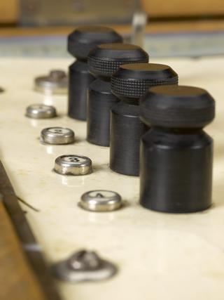
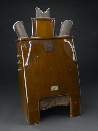
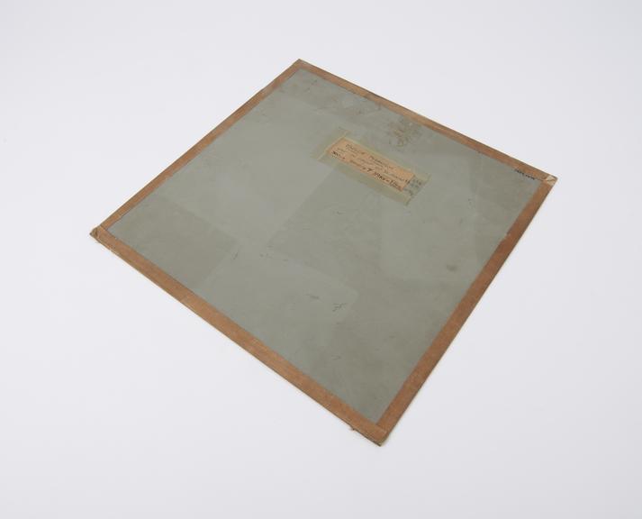
Odelca phantom, used in front of screen, marked with handwritten notes, part of Watson-Odelca mass miniature x-ray set, made by Watson and Sons (Electro-Medical), London, England, and N.V. Optische Industrie (De Oude Delft), specialist makers of X-ray equipment and optical technology, Delft, South Holland, Netherlands, used at Ilford Chest Clinic, London, England, where patients diagnosed with tuberculosis could be treated, 1960-1961
Mass miniature radiography was used to diagnose tuberculosis in Britain from 1936 onwards – in an attempt to control the disease by catching those affected as early as possible. People who appeared outwardly healthy might still show signs of tuberculosis, such as lesions in the chest, and so could spread the disease. This cream coloured machine produced X-ray images that were just 100 mm high. If physicians were unsure about a miniature X-ray when viewed on a projector, the patient was sent to hospital for a full chest X-ray.