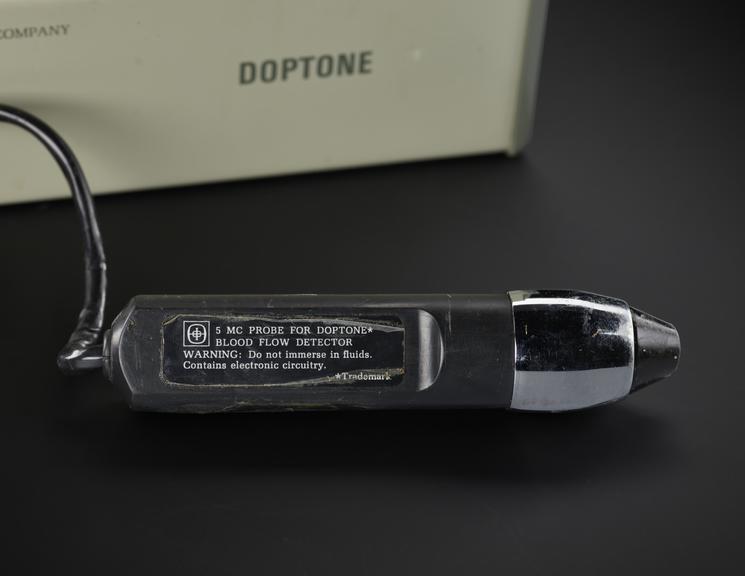
Doppler blood flow detector

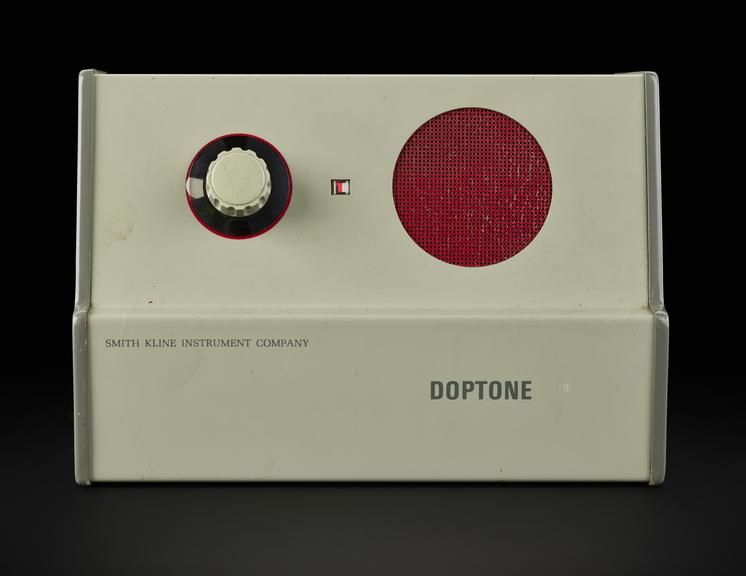
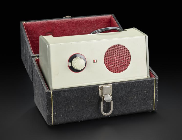
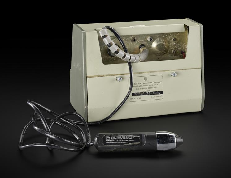
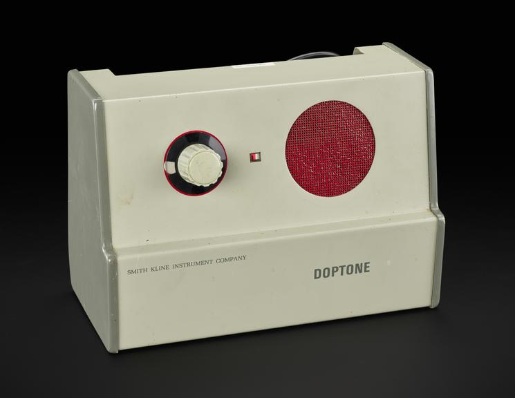
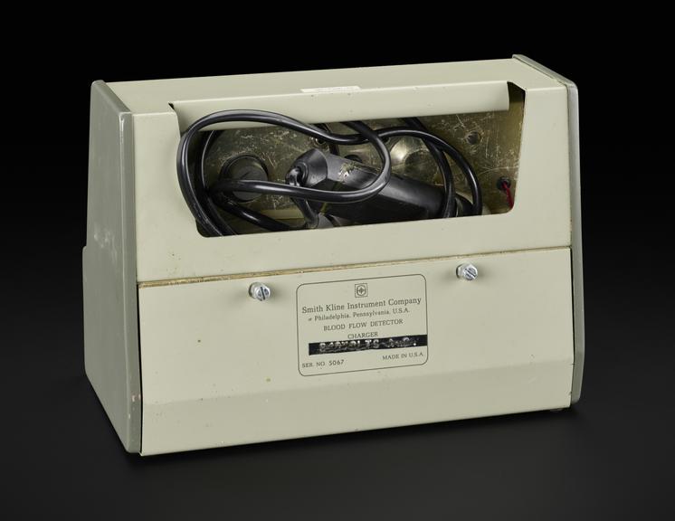
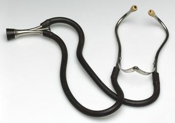
Doppler Blood flow detector, 'Doptone' with charger, probe and carrycase, serial no 5067, Smith Kline Instruments, Philadelphia, United States, 1968

First marketed in 1965, the technology behind the ‘Doptone’, was the first foetal pulse detector.

Developed at the University of Washington Cardiovascular Instrumentation Program, the machine is based on the analysis of echo signals or ‘Doppler effect’ from red blood cells in the vascular structure. The probe is applied to the skin and the user learns to listen and interpret the audio they hear. This model has a speaker and volume control to help the user. However, the audio was not enough to determine blood flow direction, the resulting sounds could not be recorded easily and there was no way of specifying how deep into the body they were scanning. In the 1970s, Doppler waves were combined with ultrasound gave user visual images to interpret and record.

Research and clinical teams realised the ‘Doptone’ could also be used to assess blood flow in the arterial and deep venous systems of the arms and legs. Peripheral vascular disease – a narrowing or blockage of a blood vessel was one area of research. Deep Vein Thrombosis (DVT), where a blood clot forms in the legs or arms, restricting blood flow can also be diagnosed through a Doppler or ultrasound.

Details

Category:
Clinical Diagnosis
Object Number:
2023-489
Materials:
metal, plastic and electronic components
Measurements:
overall: 220 mm x 280 mm x 160 mm, .05 kg
type:
ultrasound

Parts

Doppler Blood flow detector

Doppler blood flow detector, consisting of charger and probe, serial no. 5067, Smith Kline Instruments, Philadelphia, United States, 1968

More

First marketed in 1965, the technology behind the ‘Doptone’, was the first foetal pulse detector.

Developed at the University of Washington Cardiovascular Instrumentation Program, the machine is based on the analysis of echo signals or ‘Doppler effect’ from red blood cells in the vascular structure. The probe is applied to the skin and the user learns to listen and interpret the audio they hear. This model has a speaker and volume control to help the user. However, the audio was not enough to determine blood flow direction, the resulting sounds could not be recorded easily and there was no way of specifying how deep into the body they were scanning. In the 1970s, Doppler waves were combined with ultrasound gave user visual images to interpret and record.

Research and clinical teams realised the ‘Doptone’ could also be used to assess blood flow in the arterial and deep venous systems of the arms and legs. Peripheral vascular disease – a narrowing or blockage of a blood vessel was one area of research. Deep Vein Thrombosis (DVT), where a blood clot forms in the legs or arms, restricting blood flow can also be diagnosed through a Doppler or ultrasound.

Measurements:
overall: 180 mm x 255 mm x 125 mm, .05 kg
Materials:
metal , plastic and electronic components
Object Number:
2023-489/1
type:
ultrasound
Image ©
The Board of Trustees of the Science Museum
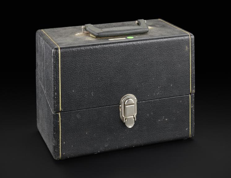
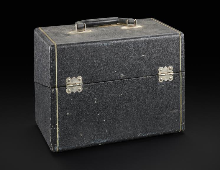
Case for Doppler blood flow detector

Fitted case for Doppler blood flow detector, Smith Kline Instruments, Philadelphia, United States, 1968

More

First marketed in 1965, the technology behind the ‘Doptone’, was the first foetal pulse detector.

Developed at the University of Washington Cardiovascular Instrumentation Program, the machine is based on the analysis of echo signals or ‘Doppler effect’ from red blood cells in the vascular structure. The probe is applied to the skin and the user learns to listen and interpret the audio they hear. This model has a speaker and volume control to help the user. However, the audio was not enough to determine blood flow direction, the resulting sounds could not be recorded easily and there was no way of specifying how deep into the body they were scanning. In the 1970s, Doppler waves were combined with ultrasound gave user visual images to interpret and record.

Research and clinical teams realised the ‘Doptone’ could also be used to assess blood flow in the arterial and deep venous systems of the arms and legs. Peripheral vascular disease – a narrowing or blockage of a blood vessel was one area of research. Deep Vein Thrombosis (DVT), where a blood clot forms in the legs or arms, restricting blood flow can also be diagnosed through a Doppler or ultrasound.

Measurements:
overall: 220 mm x 280 mm x 160 mm, .05 kg
Materials:
metal (unknown) and leather
Object Number:
2023-489/2
type:
case - container
Image ©
The Board of Trustees of the Science Museum