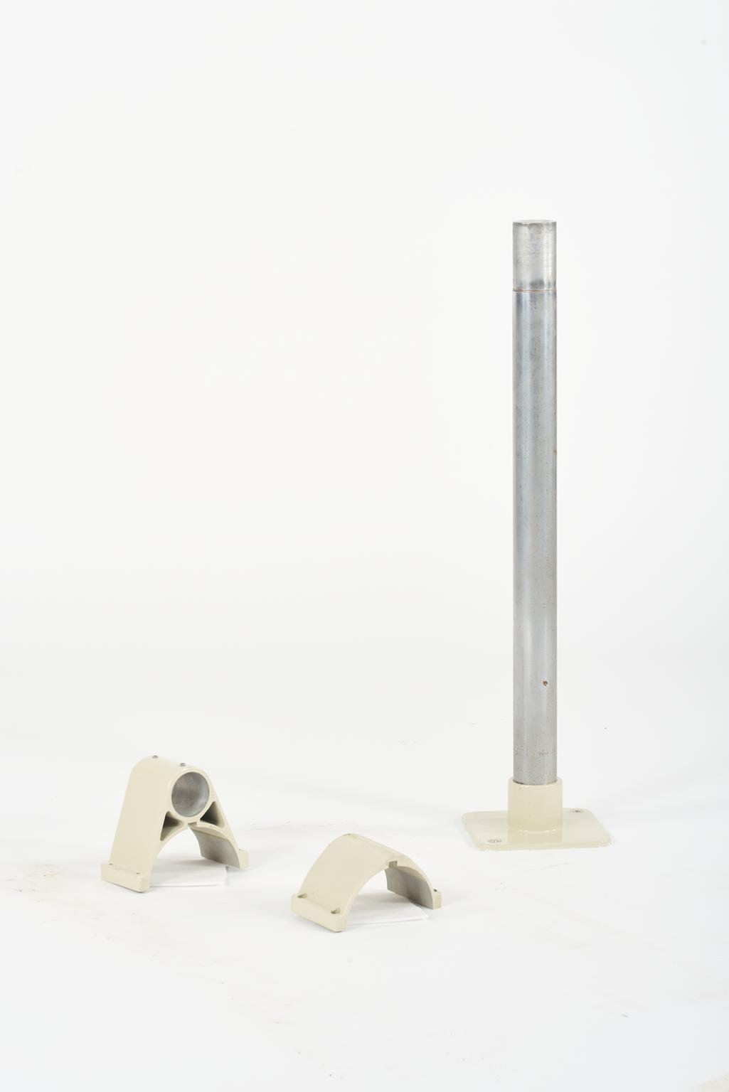
Floor to Celing Support Column for Lysholm Skull X-ray
Grey and beige floor to ceiling support column. Identification plate states property of the Queen Victoria hosp, asset number 000031. Type DS551, FAB 2615.
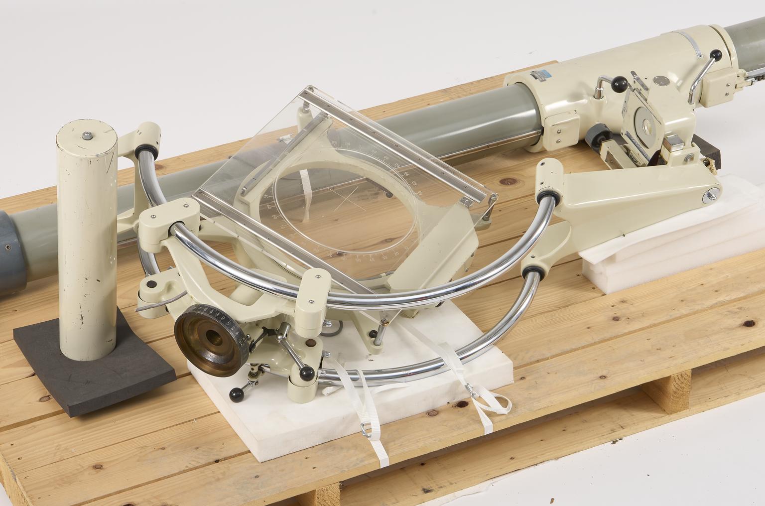
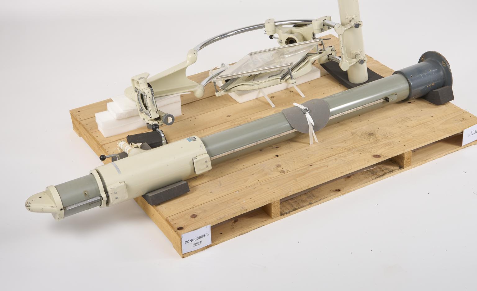
Lysholm skull x-ray imaging unit, model DS551, made by Elema-Schonander, Sweden, used for x-ray imaging facial injuries at the Queen Victoria Hospital, East Grinstead, United Kingdom which specialises in maxillofacial injuries, in use from 1940 until 2018.
More
For decades, specialist equipment like the unit shown on this slide was needed to x-ray the skull. This is one of the hardest parts of the body to image because the bone there is particularly dense and hard for x-rays to penetrate. Developed in 1931 by pioneering Swedish radiologist Erik Lysholm, the x-ray unit can rotate around the patient’s head to image it from a variety of angles, without needing to readjust the patient. It also allowed for x-rays to be taken very precisely and repeatably. For example, an x-ray could be taken of either side of the head at exactly the same angle or an x-ray could be accurately repeated on a follow up visit . This was possible because the angle of the x-ray could be very precisely set. For more than 70 years, this particular unit was used in Queen Victoria Hospital, East Grinstead – a clear demonstration of the success of its design. It was so effective that many later improvements were modelled on this unit.
The Queen Victoria Hospital’s longstanding specialism in face and jaw injuries largely dates back to the Second World War when ground-breaking surgeon Archibald McIndoe worked at the hospital. He performed pioneering plastic surgery for soldiers and airmen with facial injuries and burns.
- Measurements:
-
overall: 2400 mm x 540 mm x 240 mm,
- Materials:
- metal (unknown)
- Object Number:
- 2020-225/ 1
- type:
- support column
- Image ©
- The Board of Trustees of the Science Museum