Mallard MRI body scanner, Aberdeen, Scotland, 1983

Mallard MRI body scanner, Aberdeen, Scotland, 1983 Mallard system Magnetic Resonance Imager (MRI) body scanner by

Creative Commons LicenseThis image is released under a CC BY-NC-SA 4.0 Licence

License this image for commercial use at Science and Society Picture Library

License

Creative Commons LicenseThis image is released under a CC BY-NC-SA 4.0 Licence

License this image for commercial use at Science and Society Picture Library

License

Science Museum Group Collection
© The Board of Trustees of the Science Museum

Mallard system Magnetic Resonance Imager (MRI) body scanner by
Science Museum Group Collection
© The Board of Trustees of the Science Museum

Mallard MRI body scanner, Aberdeen, Scotland, 1983

John Mallard and his research team at Aberdeen Royal Infirmary used a machine like this in 1980 when they obtained the first clinically useful image of a patient’s internal tissues using Magnetic Resonance Imaging (MRI). Mallard’s team, based at the University of Aberdeen, was responsible for technological advances that led to the widespread introduction of MRI.

MRI is considered to be a safer diagnostic tool than X-rays and is more suitable for soft tissue. MRI builds up a picture of the human body by using high frequency radio signals. Made by M&D Technology Ltd, a company set up by John Mallard, this machine was used at St Bartholomew’s Hospital, London, from 1983 to 1993.

Details

Category:
Radiomedicine
Object Number:
1994-492
type:
mri body scanner
credit:
St Bartholomew's Hospital

Parts

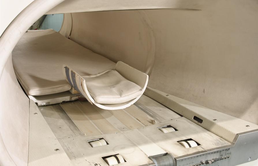
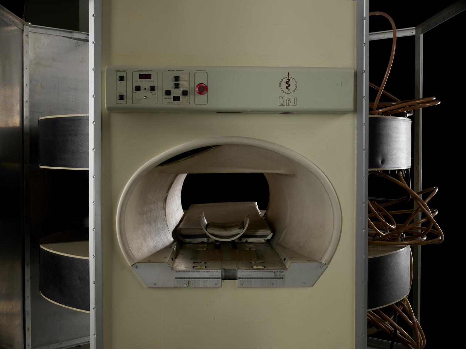
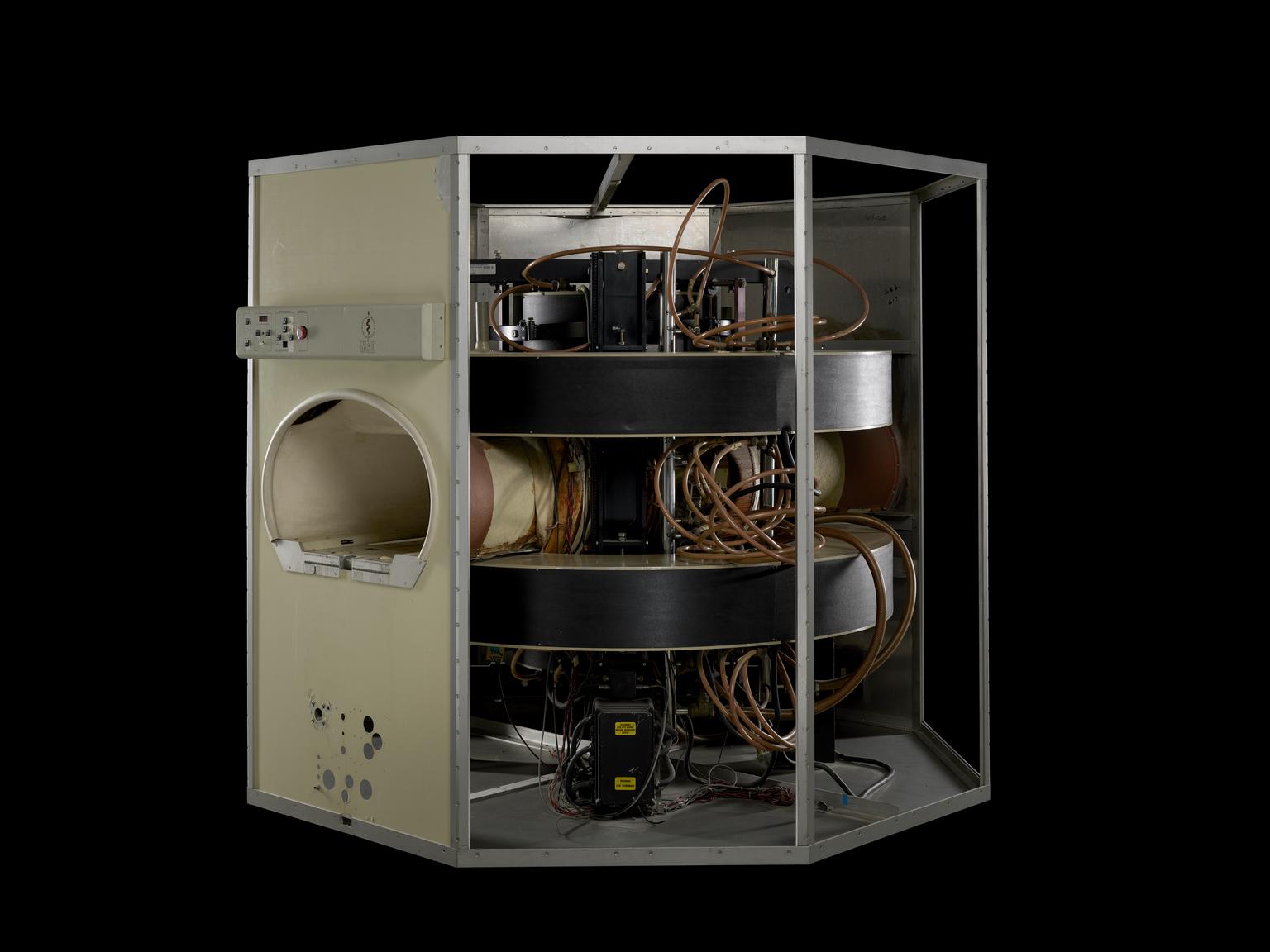
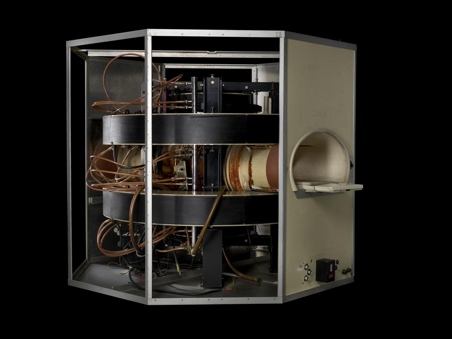
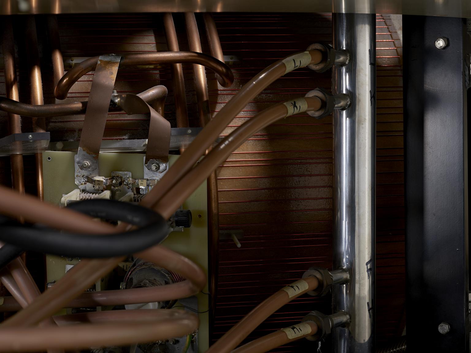
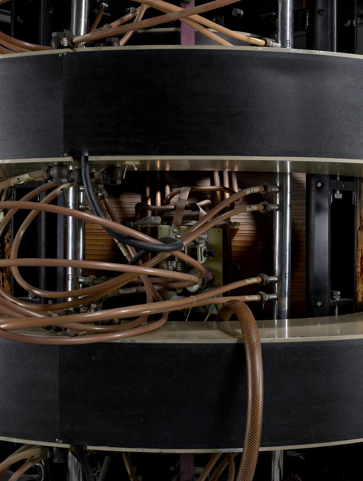
Electromagnet for Mallard MRI body scanner

Central magnet, type 621 0041, serial number 40221, consisting of a oval shaped white and brown access and patient bed, between 2 black electromagnets with attached wiring and electronic components, secured together by black steel uprights, magnet made by Oxford Instruments. Part of Mallard system Magnetic Resonance Imager (MRI) body scanner by M&D Technology, Aberdeen, 1983.

Measurements:
overall: 2400 kg
Object Number:
1994-492/1
type:
electromagnets
Image ©
The Board of Trustees of the Science Museum
Panel from Mallard MRI body scanner

Panel from Mallard MRI body scanner

Cream coloured panel with instrument panel for couch height, couch position, imaging control, emergency stop button and adhesive handwritten labels, with the maker logo, silver interior with '5' handwritten in black, and semi-circular access for patient bed, part of Mallard system Magnetic Resonance Imager (MRI) body scanner by M&D Technology, Aberdeen, 1983

Object Number:
1994-492/2
type:
panel
Panel from Mallard MRI body scanner

Panel from Mallard MRI body scanner

Cream coloured panel with silver interior with '3' handwritten in black, part of Mallard system Magnetic Resonance Imager (MRI) body scanner by M&D Technology, Aberdeen, 1983

Object Number:
1994-492/3
type:
panel
Panel from Mallard MRI body scanner

Panel from Mallard MRI body scanner

Cream coloured panel with silver interior with '4' handwritten in black, part of Mallard system Magnetic Resonance Imager (MRI) body scanner by M&D Technology, Aberdeen, 1983

Object Number:
1994-492/4
type:
panel
Panel from Mallard MRI body scanner

Panel from Mallard MRI body scanner

Cream coloured panel with semi-circular access for patient bed with silver interior with 'Inside and 486 and 417' handwritten in black, and electrical componets attached at bottom on exterio and interior, part of Mallard system Magnetic Resonance Imager (MRI) body scanner by M&D Technology, Aberdeen, 1983

Object Number:
1994-492/5
type:
panel
Frame support for Mallard MRI body scanner

Frame support for Mallard MRI body scanner

Vertical steel support for frame of Mallard system Magnetic Resonance Imager (MRI) body scanner by M&D Technology, Aberdeen, 1983. There are currently 2 screws attached to this piece

Measurements:
overall: 2013 mm x 83 mm x 40 mm,
Materials:
metal (unknown) and rubber (unidentified)
Object Number:
1994-492/6
type:
frame support
Frame Support for Mallard MRI body scanner

Frame Support for Mallard MRI body scanner

Vertical steel support for frame of Mallard system Magnetic Resonance Imager (MRI) body scanner by M&D Technology, Aberdeen, 1983. There are currently 20 screws attached to this piece

Measurements:
overall: 2013 mm x 55 mm x 28 mm,
Materials:
metal (unknown) and rubber (unidentified)
Object Number:
1994-492/7
type:
frame support
Frame Support for Mallard MRI body scanner

Frame Support for Mallard MRI body scanner

Vertical steel support for frame of Mallard system Magnetic Resonance Imager (MRI) body scanner by M&D Technology, Aberdeen, 1983. there are currently 20 screws attached to this piece.

Measurements:
overall: 2013 mm x 55 mm x 28 mm,
Materials:
metal (unknown) and rubber (unidentified)
Object Number:
1994-492/8
type:
frame support
Frame Support for Mallard MRI body scanner

Frame Support for Mallard MRI body scanner

Vertical steel support for frame of Mallard system Magnetic Resonance Imager (MRI) body scanner by M&D Technology, Aberdeen, 1983. There are currently 3 screws attached to this piece

Measurements:
overall: 2013 mm x 55 mm x 28 mm,
Materials:
metal (unknown) and rubber (unidentified)
Object Number:
1994-492/9
type:
frame support
Frame support for Mallard MRI body scanner

Frame support for Mallard MRI body scanner

Vertical steel support for frame of Mallard system Magnetic Resonance Imager (MRI) body scanner by M&D Technology, Aberdeen, 1983. There is currentyl 2 screws attached to this piece.

Measurements:
overall: 2013 mm x 55 mm x 28 mm,
Materials:
metal (unknown) and rubber (unidentified)
Object Number:
1994-492/10
type:
frame support
Frame Support for Mallard MRI body scanner

Frame Support for Mallard MRI body scanner

Vertical steel support for frame of Mallard system Magnetic Resonance Imager (MRI) body scanner by M&D Technology, Aberdeen, 1983. There are currently 2 screws attached to this piece

Measurements:
overall: 2013 mm x 83 mm x 40 mm,
Materials:
metal (unknown) and rubber (unidentified)
Object Number:
1994-492/11
type:
frame support
Frame Support for Mallard MRI body scanner

Frame Support for Mallard MRI body scanner

Vertical steel support for frame of Mallard system Magnetic Resonance Imager (MRI) body scanner by M&D Technology, Aberdeen, 1983. There are currently 2 screws attached to this piece

Measurements:
overall: 2013 mm x 83 mm x 40 mm,
Materials:
metal (unknown) and rubber (unidentified)
Object Number:
1994-492/12
type:
frame support
Frame Support for Mallard MRI body scanner

Frame Support for Mallard MRI body scanner

Vertical steel support for frame of Mallard system Magnetic Resonance Imager (MRI) body scanner by M&D Technology, Aberdeen, 1983. There are currently 2 screws attached to this piece.

Measurements:
overall: 2013 mm x 83 mm x 40 mm,
Materials:
metal (unknown) and rubber (unidentified)
Object Number:
1994-492/13
type:
frame support
Frame Support for Mallard MRI body scanner

Frame Support for Mallard MRI body scanner

Horizontal steel support for frame of Mallard system Magnetic Resonance Imager (MRI) body scanner by M&D Technology, Aberdeen, 1983. Currently has 6 screws attached to the frame support

Measurements:
overall: 1007 mm x 63 mm x 37 mm,
Materials:
metal (unknown) and rubber (unidentified)
Object Number:
1994-492/14
type:
frame support
Frame support for Mallard MRI body scanner

Frame support for Mallard MRI body scanner

Horizontal steel support for frame of Mallard system Magnetic Resonance Imager (MRI) body scanner by M&D Technology, Aberdeen, 1983. There are currently 7 screws attached to the frame support

Measurements:
overall: 1013 mm x 94 mm x 50 mm,
Materials:
metal (unknown)
Object Number:
1994-492/15
type:
frame support
Frame support for Mallard MRI body scanner

Frame support for Mallard MRI body scanner

Horizontal steel support for frame of Mallard system Magnetic Resonance Imager (MRI) body scanner by M&D Technology, Aberdeen, 1983

Measurements:
overall: 1007 mm x 50 mm x 7 mm,
Materials:
metal (unknown)
Object Number:
1994-492/16
type:
frame support
Frame Support for Mallard MRI body scanner

Frame Support for Mallard MRI body scanner

Horizontal steel support for frame of Mallard system Magnetic Resonance Imager (MRI) body scanner by M&D Technology, Aberdeen, 1983. This part currently has 9 screws attached to the frame support

Measurements:
overall: 1015 mm x 45 mm x 50 mm,
Materials:
metal (unknown) and rubber (unidentified)
Object Number:
1994-492/17
type:
frame support
Frame support for Mallard MRI body scanner

Frame support for Mallard MRI body scanner

Horizontal steel support for frame of Mallard system Magnetic Resonance Imager (MRI) body scanner by M&D Technology, Aberdeen, 1983

Measurements:
overall: 1007 mm x 50 mm x 7 mm,
Materials:
metal (unknown)
Object Number:
1994-492/18
type:
frame support
Frame support for Mallard MRI body scanner

Frame support for Mallard MRI body scanner

Horizontal steel support for frame of Mallard system Magnetic Resonance Imager (MRI) body scanner by M&D Technology, Aberdeen, 1983

Measurements:
overall: 1007 mm x 50 mm x 7 mm,
Materials:
metal (unknown)
Object Number:
1994-492/19
type:
frame support
Frame support for Mallard MRI body scanner

Frame support for Mallard MRI body scanner

Horizontal steel support for frame of Mallard system Magnetic Resonance Imager (MRI) body scanner by M&D Technology, Aberdeen, 1983

Measurements:
overall: 1007 mm x 50 mm x 7 mm,
Materials:
metal (unknown)
Object Number:
1994-492/20
type:
frame support
Metal Pole Wire Cover from Mallard MRI body scanner

Metal Pole Wire Cover from Mallard MRI body scanner

Metal pole wire cover, part of Mallard system Magnetic Resonance Imager (MRI) body scanner by M&D Technology, Aberdeen, 1983

More

John Mallard and his research team at Aberdeen Royal Infirmary used a machine like this in 1980 when they obtained the first clinically useful image of a patient’s internal tissues using Magnetic Resonance Imaging (MRI). Mallard’s team, based at the University of Aberdeen, was responsible for technological advances that led to the widespread introduction of MRI.

MRI is considered to be a safer diagnostic tool than X-rays and is more suitable for soft tissue. MRI builds up a picture of the human body by using high frequency radio signals. Made by M&D Technology Ltd, a company set up by John Mallard, this machine was used at St Bartholomew’s Hospital, London, from 1983 to 1993.

Measurements:
overall: 1730 mm x 260 mm x 90 mm,
Materials:
metal (unknown) and rubber (unidentified)
Object Number:
1994-492/21
type:
metal pole wire cover
Frame support for Mallard MRI body scanner

Frame support for Mallard MRI body scanner

Horizontal steel support for frame of Mallard system Magnetic Resonance Imager (MRI) body scanner by M&D Technology, Aberdeen, 1983

Measurements:
overall: 1007 mm x 50 mm x 7 mm,
Materials:
metal (unknown)
Object Number:
1994-492/22
type:
frame support
Frame support for Mallard MRI body scanner

Frame support for Mallard MRI body scanner

Horizontal steel support for frame of Mallard system Magnetic Resonance Imager (MRI) body scanner by M&D Technology, Aberdeen, 1983. Currently has 12 screws attached to the frame support

Measurements:
overall: 1007 mm x 93 mm x 50 mm,
Materials:
metal (unknown) and rubber (unidentified)
Object Number:
1994-492/23
type:
frame support
Frame support for Mallard MRI body scanner

Frame support for Mallard MRI body scanner

Horizontal steel support for frame of Mallard system Magnetic Resonance Imager (MRI) body scanner by M&D Technology, Aberdeen, 1983. There are 8 screws currently attached to the frame.

Measurements:
overall: 1015 mm x 48 mm x 50 mm,
Materials:
metal (unknown)
Object Number:
1994-492/24
type:
frame support
Frame support for Mallard MRI body scanner

Frame support for Mallard MRI body scanner

Horizontal steel support for frame of Mallard system Magnetic Resonance Imager (MRI) body scanner by M&D Technology, Aberdeen, 1983. Currently has 7 screws attached to the frame support.

A bag with rubber gaskets has been attached to this frame support piece.

Measurements:
overall: 115 mm x 65 mm x 37 mm,
Materials:
metal (unknown)
Object Number:
1994-492/25
type:
frame support
Frame support for Mallard MRI body scanner

Frame support for Mallard MRI body scanner

Horizontal steel support for frame of Mallard system Magnetic Resonance Imager (MRI) body scanner by M&D Technology, Aberdeen, 1983

Measurements:
overall: 1007 mm x 50 mm x 7 mm,
Materials:
metal (unknown)
Object Number:
1994-492/26
type:
frame support
Frame support for Mallard MRI body scanner

Frame support for Mallard MRI body scanner

Horizontal steel support for frame of Mallard system Magnetic Resonance Imager (MRI) body scanner by M&D Technology, Aberdeen, 1983. Currently has 3 screws attached to the frame support

Measurements:
overall: 1030 mm x 48 mm x 50 mm,
Materials:
metal (unknown) and rubber (unidentified)
Object Number:
1994-492/27
type:
frame support
Frame Support for Mallard MRI body scanner

Frame Support for Mallard MRI body scanner

Horizontal steel support for frame of Mallard system Magnetic Resonance Imager (MRI) body scanner by M&D Technology, Aberdeen, 1983. There are currently 6 screws attached to this frame support piece.

Measurements:
overall: 1015 mm x 50 mm x 45 mm,
Materials:
metal (unknown) and rubber (unidentified)
Object Number:
1994-492/28
type:
frame support
Frame support for Mallard MRI body scanner

Frame support for Mallard MRI body scanner

Horizontal steel support for frame of Mallard system Magnetic Resonance Imager (MRI) body scanner by M&D Technology, Aberdeen, 1983

Measurements:
overall: 1013 mm x 50 mm x 6 mm,
Materials:
metal (unknown)
Object Number:
1994-492/29
type:
frame support
Frame support for Mallard MRI body scanner

Frame support for Mallard MRI body scanner

Horizontal steel support for frame of Mallard system Magnetic Resonance Imager (MRI) body scanner by M&D Technology, Aberdeen, 1983

Measurements:
overall: 1007 mm x 50 mm x 7 mm,
Materials:
metal (unknown)
Object Number:
1994-492/30
type:
frame support
Top Frame Support for Mallard MRI body scanner

Top Frame Support for Mallard MRI body scanner

Steel support with middle joint for the width of the frame of Mallard system Magnetic Resonance Imager (MRI) body scanner by M&D Technology, Aberdeen, 1983, There are currently 5 screws - 2 of which are broken - attached to this piece

Measurements:
overall: 2355 mm x 50 mm x 63 mm,
Materials:
metal (unknown)
Object Number:
1994-492/31
type:
frame support
Base Plate from Mallard MRI body scanner

Base Plate from Mallard MRI body scanner

Semi-octagonal steel base plate, part of Mallard system Magnetic Resonance Imager (MRI) body scanner by M&D Technology, Aberdeen, 1983

Measurements:
overall: 2430 mm x 2430 mm x 65 mm,
Materials:
metal (unknown) , textile and plastic (unidentified)
Object Number:
1994-492/32
type:
base plate
Control Panel Unit from Mallard MRI body scanner

Control Panel Unit from Mallard MRI body scanner

Control panel unit, part of Mallard system Magnetic Resonance Imager (MRI) body scanner by M&D Technology, Aberdeen, 1983. There is currently a bag attached to this piece. the contents of the bag go inside the control panel when being rebuilt in the future. There are currently 26 visible screws attached to this piece.

Measurements:
overall: 900 mm x 165 mm x 110 mm,
Materials:
metal (unknown) , plastic (unidentified) and rubber (unidentified)
Object Number:
1994-492/33
type:
control panel
Screws from Mallard MRI body scanner

Screws from Mallard MRI body scanner

Screws from semi-circular access for patient bed, part of Mallard system Magnetic Resonance Imager (MRI) body scanner by M&D Technology, Aberdeen, 1983

Object Number:
1994-492/34
type:
screws
Screws from Mallard MRI body scanner

Screws from Mallard MRI body scanner

Screws from semi-circular access for patient bed, part of Mallard system Magnetic Resonance Imager (MRI) body scanner by M&D Technology, Aberdeen, 1983

Object Number:
1994-492/35
type:
screws
Patient's Rolling Bed from Mallard MRI body scanner

Patient's Rolling Bed from Mallard MRI body scanner

Patient's rolling bed, part of Mallard system Magnetic Resonance Imager (MRI) body scanner by M&D Technology, Aberdeen, 1983

Object Number:
1994-492/36
type:
bed
Bed Roller Extension from Mallard MRI body scanner

Bed Roller Extension from Mallard MRI body scanner

Bed Roller Extension, part of Mallard system Magnetic Resonance Imager (MRI) body scanner by M&D Technology, Aberdeen, 1983.

More

John Mallard and his research team at Aberdeen Royal Infirmary used a machine like this in 1980 when they obtained the first clinically useful image of a patient’s internal tissues using Magnetic Resonance Imaging (MRI). Mallard’s team, based at the University of Aberdeen, was responsible for technological advances that led to the widespread introduction of MRI.

MRI is considered to be a safer diagnostic tool than X-rays and is more suitable for soft tissue. MRI builds up a picture of the human body by using high frequency radio signals. Made by M&D Technology Ltd, a company set up by John Mallard, this machine was used at St Bartholomew’s Hospital, London, from 1983 to 1993.

Measurements:
overall: 990 mm x 582 mm x 245 mm,
Materials:
metal (unknown) , plastic (unidentified) and rubber (unidentified)
Object Number:
1994-492/37
type:
bed
Frame and Rubber Gasket from the Semi-Circular Access for Patient Bed from Mallard MRI Body Scanner

Frame and Rubber Gasket from the Semi-Circular Access for Patient Bed from Mallard MRI Body Scanner

Frame and rubber gasket from the semi-circular access for patient bed, part of Mallard system Magnetic Resonance Imager (MRI) body scanner by M&D Technology, Aberdeen, 1983

Object Number:
1994-492/38
type:
frame support
Frame and Rubber Gasket From the Semi-circular Opening on the Control Panel End From Mallard MRI body scanner

Frame and Rubber Gasket From the Semi-circular Opening on the Control Panel End From Mallard MRI body scanner

Frame and rubber gasket from the semi-circular opening on the control panel end, part of Mallard system Magnetic Resonance Imager (MRI) body scanner by M&D Technology, Aberdeen, 1983.

More

John Mallard and his research team at Aberdeen Royal Infirmary used a machine like this in 1980 when they obtained the first clinically useful image of a patient’s internal tissues using Magnetic Resonance Imaging (MRI). Mallard’s team, based at the University of Aberdeen, was responsible for technological advances that led to the widespread introduction of MRI.

MRI is considered to be a safer diagnostic tool than X-rays and is more suitable for soft tissue. MRI builds up a picture of the human body by using high frequency radio signals. Made by M&D Technology Ltd, a company set up by John Mallard, this machine was used at St Bartholomew’s Hospital, London, from 1983 to 1993.

Object Number:
1994-492/39
type:
frame support
Faceplate (right) from Mallard MRI Body Scanner

Faceplate (right) from Mallard MRI Body Scanner

Faceplate (right), part of Mallard system Magnetic Resonance Imager (MRI) body scanner by M&D Technology, Aberdeen, 1983

Measurements:
overall: 340 mm x 93 mm x 12 mm,
Materials:
metal (unknown)
Object Number:
1994-492/40
type:
faceplate
Faceplate (left) from Mallard MRI Body Scanner

Faceplate (left) from Mallard MRI Body Scanner

Faceplate (left), part of Mallard system Magnetic Resonance Imager (MRI) body scanner by M&D Technology, Aberdeen, 1983.

More

John Mallard and his research team at Aberdeen Royal Infirmary used a machine like this in 1980 when they obtained the first clinically useful image of a patient’s internal tissues using Magnetic Resonance Imaging (MRI). Mallard’s team, based at the University of Aberdeen, was responsible for technological advances that led to the widespread introduction of MRI.

MRI is considered to be a safer diagnostic tool than X-rays and is more suitable for soft tissue. MRI builds up a picture of the human body by using high frequency radio signals. Made by M&D Technology Ltd, a company set up by John Mallard, this machine was used at St Bartholomew’s Hospital, London, from 1983 to 1993.

Measurements:
overall: 340 mm x 93 mm x 12 mm,
Materials:
metal (unknown)
Object Number:
1994-492/41
type:
faceplate
Faceplate from Mallard MRI Body Scanner

Faceplate from Mallard MRI Body Scanner

Faceplate, part of Mallard system Magnetic Resonance Imager (MRI) body scanner by M&D Technology, Aberdeen, 1983

Measurements:
overall: 740 mm x 68 mm x 13 mm,
Materials:
metal (unknown)
Object Number:
1994-492/42
type:
faceplate
Horizontal Frame Support Above Bed from Mallard MRI Body Scanner

Horizontal Frame Support Above Bed from Mallard MRI Body Scanner

Horizontal Frame Support from above bed, part of Mallard system Magnetic Resonance Imager (MRI) body scanner by M&D Technology, Aberdeen, 1983.

More

John Mallard and his research team at Aberdeen Royal Infirmary used a machine like this in 1980 when they obtained the first clinically useful image of a patient’s internal tissues using Magnetic Resonance Imaging (MRI). Mallard’s team, based at the University of Aberdeen, was responsible for technological advances that led to the widespread introduction of MRI.

MRI is considered to be a safer diagnostic tool than X-rays and is more suitable for soft tissue. MRI builds up a picture of the human body by using high frequency radio signals. Made by M&D Technology Ltd, a company set up by John Mallard, this machine was used at St Bartholomew’s Hospital, London, from 1983 to 1993.

Measurements:
overall: 993 mm x 50 mm x 50 mm,
Materials:
metal (unknown) and rubber (unidentified)
Object Number:
1994-492/43
type:
frame support
Horizontal Frame Support Below Bed from Mallard MRI Body Scanner

Horizontal Frame Support Below Bed from Mallard MRI Body Scanner

Horizontal frame support for below bed, part of Mallard system Magnetic Resonance Imager (MRI) body scanner by M&D Technology, Aberdeen, 1983. Currently has 2 screws attached to the frame support

Measurements:
overall: 938 mm x 63 mm x 63 mm,
Materials:
metal (unknown)
Object Number:
1994-492/44
type:
frame support
Horizontal Frame Support Above Bed from Mallard MRI Body Scanner

Horizontal Frame Support Above Bed from Mallard MRI Body Scanner

Horizontal Frame Support from above the bed, part of Mallard system Magnetic Resonance Imager (MRI) body scanner by M&D Technology, Aberdeen, 1983.

More

John Mallard and his research team at Aberdeen Royal Infirmary used a machine like this in 1980 when they obtained the first clinically useful image of a patient’s internal tissues using Magnetic Resonance Imaging (MRI). Mallard’s team, based at the University of Aberdeen, was responsible for technological advances that led to the widespread introduction of MRI.

MRI is considered to be a safer diagnostic tool than X-rays and is more suitable for soft tissue. MRI builds up a picture of the human body by using high frequency radio signals. Made by M&D Technology Ltd, a company set up by John Mallard, this machine was used at St Bartholomew’s Hospital, London, from 1983 to 1993.

Measurements:
overall: 995 mm x 50 mm x 50 mm,
Materials:
metal (unknown)
Object Number:
1994-492/45
type:
frame support
Horizontal Frame Support Below Bed from Mallard MRI Body Scanner

Horizontal Frame Support Below Bed from Mallard MRI Body Scanner

Horizontal frame support below bed, part of Mallard system Magnetic Resonance Imager (MRI) body scanner by M&D Technology, Aberdeen, 1983

Measurements:
overall: 940 mm x 60 mm x 63 mm,
Materials:
metal (unknown) and plastic (unidentified)
Object Number:
1994-492/46
type:
frame support