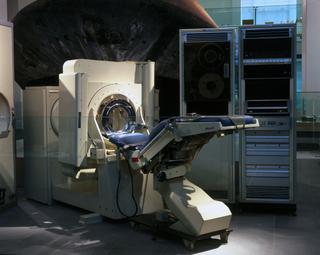
Reynolds' cineradiography apparatus

16mm cineradiography set, Dr. R J Reynold's design, comprises grid-enclosed power-supply, tube stand, x-ray tube, rectifier, screening stand, patient stand, camera unit, control unit and accessories by Watson and Sons (Electro-Medical) Ltd, London, 1935

Russell John Reynolds was an internationally renowned radiographer and specialist in the field of cineradiography (moving image X-ray films). While still at school, he – with the assistance of his GP father and physicist Sir William Crookes – constructed a fully functioning X-ray machine just months after German scientist Wilhelm Röntgen first described the ‘new type of ray’ in late 1895.

Qualifying as a doctor in 1907, he spent two years in general practice before becoming a full-time radiologist, going on to serve with the Royal Army Medical Corps during World War I. In 1921, he was appointed physician-in-charge of the X-ray departments of Charing Cross and the National Hospitals where he began tackling the challenge of creating X-ray films of moving body parts.

At the time, there were two possible ways of producing cineradiographic films. The ‘direct’ method, as the name suggests, recorded X-ray images directly onto cinefilm. As X-rays could not be focused, film strips had to be as wide as the body part being X-rayed. To capture adult chest movements, for example, a reel 15 inches wide by 120 feet long would be needed.

Reynolds favoured the ‘indirect’ approach, also known as cinefluorography: recording live X-ray images from a fluorescent screen. This tactic presented its own challenges, namely producing a bright enough image to show up on film without exposing the patient to harmful levels of radiation. Reynolds constantly modified his techniques to reduce the exposure times needed. By 1925, he had introduced a switch to automatically cut off the X-ray tube while the camera shutter was closed.

Within a decade, Reynolds had filmed almost all the body’s joints at speeds of up to 12 frames per second and persuaded optical instrument maker W. Watson and Sons to develop the first commercial cineradiography machine. Cautious about profitability, the company adapted existing equipment to create the outfit – the camera base, for example, is a modified dental chair. Reynolds continued to experiment in subsequent decades, achieving films of 50 frames per second, ‘slow motion’ imagery of the heartbeat and barium meal examinations of the intestines.

Parts

Control unit for cineradiography set

Control unit for cineradiography set

Control unit for cineradiography set

More

Russell John Reynolds was an internationally renowned radiographer and specialist in the field of cineradiography (moving image X-ray films). While still at school, he – with the assistance of his GP father and physicist Sir William Crookes – constructed a fully functioning X-ray machine just months after German scientist Wilhelm Röntgen first described the ‘new type of ray’ in late 1895.

Qualifying as a doctor in 1907, he spent two years in general practice before becoming a full-time radiologist, going on to serve with the Royal Army Medical Corps during World War I. In 1921, he was appointed physician-in-charge of the X-ray departments of Charing Cross and the National Hospitals where he began tackling the challenge of creating X-ray films of moving body parts.

At the time, there were two possible ways of producing cineradiographic films. The ‘direct’ method, as the name suggests, recorded X-ray images directly onto cinefilm. As X-rays could not be focused, film strips had to be as wide as the body part being X-rayed. To capture adult chest movements, for example, a reel 15 inches wide by 120 feet long would be needed.

Reynolds favoured the ‘indirect’ approach, also known as cinefluorography: recording live X-ray images from a fluorescent screen. This tactic presented its own challenges, namely producing a bright enough image to show up on film without exposing the patient to harmful levels of radiation. Reynolds constantly modified his techniques to reduce the exposure times needed. By 1925, he had introduced a switch to automatically cut off the X-ray tube while the camera shutter was closed.

Within a decade, Reynolds had filmed almost all the body’s joints at speeds of up to 12 frames per second and persuaded optical instrument maker W. Watson and Sons to develop the first commercial cineradiography machine. Cautious about profitability, the company adapted existing equipment to create the outfit – the camera base, for example, is a modified dental chair. Reynolds continued to experiment in subsequent decades, achieving films of 50 frames per second, ‘slow motion’ imagery of the heartbeat and barium meal examinations of the intestines.

Measurements:
overall: 937 mm x 450 mm x 429 mm, 102 kg
Materials:
iron and steel
Object Number:
A639410 Pt1
type:
radioactive material and control panels
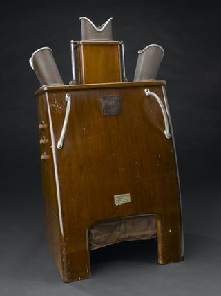
Cineradiography equipment

Cineradiography equipment

High tension supply for cineradiography set in mesh safety enclosure also holding "telestat" x-ray tube stand.

More

Russell John Reynolds was an internationally renowned radiographer and specialist in the field of cineradiography (moving image X-ray films). While still at school, he – with the assistance of his GP father and physicist Sir William Crookes – constructed a fully functioning X-ray machine just months after German scientist Wilhelm Röntgen first described the ‘new type of ray’ in late 1895.

Qualifying as a doctor in 1907, he spent two years in general practice before becoming a full-time radiologist, going on to serve with the Royal Army Medical Corps during World War I. In 1921, he was appointed physician-in-charge of the X-ray departments of Charing Cross and the National Hospitals where he began tackling the challenge of creating X-ray films of moving body parts.

At the time, there were two possible ways of producing cineradiographic films. The ‘direct’ method, as the name suggests, recorded X-ray images directly onto cinefilm. As X-rays could not be focused, film strips had to be as wide as the body part being X-rayed. To capture adult chest movements, for example, a reel 15 inches wide by 120 feet long would be needed.

Reynolds favoured the ‘indirect’ approach, also known as cinefluorography: recording live X-ray images from a fluorescent screen. This tactic presented its own challenges, namely producing a bright enough image to show up on film without exposing the patient to harmful levels of radiation. Reynolds constantly modified his techniques to reduce the exposure times needed. By 1925, he had introduced a switch to automatically cut off the X-ray tube while the camera shutter was closed.

Within a decade, Reynolds had filmed almost all the body’s joints at speeds of up to 12 frames per second and persuaded optical instrument maker W. Watson and Sons to develop the first commercial cineradiography machine. Cautious about profitability, the company adapted existing equipment to create the outfit – the camera base, for example, is a modified dental chair. Reynolds continued to experiment in subsequent decades, achieving films of 50 frames per second, ‘slow motion’ imagery of the heartbeat and barium meal examinations of the intestines.

Metalix x-ray tube

Metalix x-ray tube

"Metalix" x-ray tube type 20631 with heatsink for air-cooling, by Philips, Holland, 1935-1945

More

Russell John Reynolds was an internationally renowned radiographer and specialist in the field of cineradiography (moving image X-ray films). While still at school, he – with the assistance of his GP father and physicist Sir William Crookes – constructed a fully functioning X-ray machine just months after German scientist Wilhelm Röntgen first described the ‘new type of ray’ in late 1895.

Qualifying as a doctor in 1907, he spent two years in general practice before becoming a full-time radiologist, going on to serve with the Royal Army Medical Corps during World War I. In 1921, he was appointed physician-in-charge of the X-ray departments of Charing Cross and the National Hospitals where he began tackling the challenge of creating X-ray films of moving body parts.

At the time, there were two possible ways of producing cineradiographic films. The ‘direct’ method, as the name suggests, recorded X-ray images directly onto cinefilm. As X-rays could not be focused, film strips had to be as wide as the body part being X-rayed. To capture adult chest movements, for example, a reel 15 inches wide by 120 feet long would be needed.

Reynolds favoured the ‘indirect’ approach, also known as cinefluorography: recording live X-ray images from a fluorescent screen. This tactic presented its own challenges, namely producing a bright enough image to show up on film without exposing the patient to harmful levels of radiation. Reynolds constantly modified his techniques to reduce the exposure times needed. By 1925, he had introduced a switch to automatically cut off the X-ray tube while the camera shutter was closed.

Within a decade, Reynolds had filmed almost all the body’s joints at speeds of up to 12 frames per second and persuaded optical instrument maker W. Watson and Sons to develop the first commercial cineradiography machine. Cautious about profitability, the company adapted existing equipment to create the outfit – the camera base, for example, is a modified dental chair. Reynolds continued to experiment in subsequent decades, achieving films of 50 frames per second, ‘slow motion’ imagery of the heartbeat and barium meal examinations of the intestines.

Materials:
steel and glass
Object Number:
A639410 Pt3
type:
x-ray tubes
x-ray rectifier tubes

x-ray rectifier tubes

Thermionic rectifier tube, type 28201, thought to be made by Philips, 1935-1950

More

Russell John Reynolds was an internationally renowned radiographer and specialist in the field of cineradiography (moving image X-ray films). While still at school, he – with the assistance of his GP father and physicist Sir William Crookes – constructed a fully functioning X-ray machine just months after German scientist Wilhelm Röntgen first described the ‘new type of ray’ in late 1895.

Qualifying as a doctor in 1907, he spent two years in general practice before becoming a full-time radiologist, going on to serve with the Royal Army Medical Corps during World War I. In 1921, he was appointed physician-in-charge of the X-ray departments of Charing Cross and the National Hospitals where he began tackling the challenge of creating X-ray films of moving body parts.

At the time, there were two possible ways of producing cineradiographic films. The ‘direct’ method, as the name suggests, recorded X-ray images directly onto cinefilm. As X-rays could not be focused, film strips had to be as wide as the body part being X-rayed. To capture adult chest movements, for example, a reel 15 inches wide by 120 feet long would be needed.

Reynolds favoured the ‘indirect’ approach, also known as cinefluorography: recording live X-ray images from a fluorescent screen. This tactic presented its own challenges, namely producing a bright enough image to show up on film without exposing the patient to harmful levels of radiation. Reynolds constantly modified his techniques to reduce the exposure times needed. By 1925, he had introduced a switch to automatically cut off the X-ray tube while the camera shutter was closed.

Within a decade, Reynolds had filmed almost all the body’s joints at speeds of up to 12 frames per second and persuaded optical instrument maker W. Watson and Sons to develop the first commercial cineradiography machine. Cautious about profitability, the company adapted existing equipment to create the outfit – the camera base, for example, is a modified dental chair. Reynolds continued to experiment in subsequent decades, achieving films of 50 frames per second, ‘slow motion’ imagery of the heartbeat and barium meal examinations of the intestines.

Measurements:
overall: 450 mm 110 mm,
Materials:
chrome steel and plastic
Object Number:
A639410 Pt4
type:
tube
Backboard for x-ray machine

Backboard for x-ray machine

Backboard for x-ray machine

More

Russell John Reynolds was an internationally renowned radiographer and specialist in the field of cineradiography (moving image X-ray films). While still at school, he – with the assistance of his GP father and physicist Sir William Crookes – constructed a fully functioning X-ray machine just months after German scientist Wilhelm Röntgen first described the ‘new type of ray’ in late 1895.

Qualifying as a doctor in 1907, he spent two years in general practice before becoming a full-time radiologist, going on to serve with the Royal Army Medical Corps during World War I. In 1921, he was appointed physician-in-charge of the X-ray departments of Charing Cross and the National Hospitals where he began tackling the challenge of creating X-ray films of moving body parts.

At the time, there were two possible ways of producing cineradiographic films. The ‘direct’ method, as the name suggests, recorded X-ray images directly onto cinefilm. As X-rays could not be focused, film strips had to be as wide as the body part being X-rayed. To capture adult chest movements, for example, a reel 15 inches wide by 120 feet long would be needed.

Reynolds favoured the ‘indirect’ approach, also known as cinefluorography: recording live X-ray images from a fluorescent screen. This tactic presented its own challenges, namely producing a bright enough image to show up on film without exposing the patient to harmful levels of radiation. Reynolds constantly modified his techniques to reduce the exposure times needed. By 1925, he had introduced a switch to automatically cut off the X-ray tube while the camera shutter was closed.

Within a decade, Reynolds had filmed almost all the body’s joints at speeds of up to 12 frames per second and persuaded optical instrument maker W. Watson and Sons to develop the first commercial cineradiography machine. Cautious about profitability, the company adapted existing equipment to create the outfit – the camera base, for example, is a modified dental chair. Reynolds continued to experiment in subsequent decades, achieving films of 50 frames per second, ‘slow motion’ imagery of the heartbeat and barium meal examinations of the intestines.

Vertical fluorescent screening stand

Vertical fluorescent screening stand

Vertical fluorescent screening stand, wheeled, part of cineradiography apparatus

More

Russell John Reynolds was an internationally renowned radiographer and specialist in the field of cineradiography (moving image X-ray films). While still at school, he – with the assistance of his GP father and physicist Sir William Crookes – constructed a fully functioning X-ray machine just months after German scientist Wilhelm Röntgen first described the ‘new type of ray’ in late 1895.

Qualifying as a doctor in 1907, he spent two years in general practice before becoming a full-time radiologist, going on to serve with the Royal Army Medical Corps during World War I. In 1921, he was appointed physician-in-charge of the X-ray departments of Charing Cross and the National Hospitals where he began tackling the challenge of creating X-ray films of moving body parts.

At the time, there were two possible ways of producing cineradiographic films. The ‘direct’ method, as the name suggests, recorded X-ray images directly onto cinefilm. As X-rays could not be focused, film strips had to be as wide as the body part being X-rayed. To capture adult chest movements, for example, a reel 15 inches wide by 120 feet long would be needed.

Reynolds favoured the ‘indirect’ approach, also known as cinefluorography: recording live X-ray images from a fluorescent screen. This tactic presented its own challenges, namely producing a bright enough image to show up on film without exposing the patient to harmful levels of radiation. Reynolds constantly modified his techniques to reduce the exposure times needed. By 1925, he had introduced a switch to automatically cut off the X-ray tube while the camera shutter was closed.

Within a decade, Reynolds had filmed almost all the body’s joints at speeds of up to 12 frames per second and persuaded optical instrument maker W. Watson and Sons to develop the first commercial cineradiography machine. Cautious about profitability, the company adapted existing equipment to create the outfit – the camera base, for example, is a modified dental chair. Reynolds continued to experiment in subsequent decades, achieving films of 50 frames per second, ‘slow motion’ imagery of the heartbeat and barium meal examinations of the intestines.

Materials:
iron , steel and window, glass
Object Number:
A639410 Pt6
type:
protective screens
Camera and synchronisation unit for cineradiography set (camera missing)

Camera and synchronisation unit for cineradiography set (camera missing)

Camera and synchronisation unit for cineradiography set (camera missing)

More

Russell John Reynolds was an internationally renowned radiographer and specialist in the field of cineradiography (moving image X-ray films). While still at school, he – with the assistance of his GP father and physicist Sir William Crookes – constructed a fully functioning X-ray machine just months after German scientist Wilhelm Röntgen first described the ‘new type of ray’ in late 1895.

Qualifying as a doctor in 1907, he spent two years in general practice before becoming a full-time radiologist, going on to serve with the Royal Army Medical Corps during World War I. In 1921, he was appointed physician-in-charge of the X-ray departments of Charing Cross and the National Hospitals where he began tackling the challenge of creating X-ray films of moving body parts.

At the time, there were two possible ways of producing cineradiographic films. The ‘direct’ method, as the name suggests, recorded X-ray images directly onto cinefilm. As X-rays could not be focused, film strips had to be as wide as the body part being X-rayed. To capture adult chest movements, for example, a reel 15 inches wide by 120 feet long would be needed.

Reynolds favoured the ‘indirect’ approach, also known as cinefluorography: recording live X-ray images from a fluorescent screen. This tactic presented its own challenges, namely producing a bright enough image to show up on film without exposing the patient to harmful levels of radiation. Reynolds constantly modified his techniques to reduce the exposure times needed. By 1925, he had introduced a switch to automatically cut off the X-ray tube while the camera shutter was closed.

Within a decade, Reynolds had filmed almost all the body’s joints at speeds of up to 12 frames per second and persuaded optical instrument maker W. Watson and Sons to develop the first commercial cineradiography machine. Cautious about profitability, the company adapted existing equipment to create the outfit – the camera base, for example, is a modified dental chair. Reynolds continued to experiment in subsequent decades, achieving films of 50 frames per second, ‘slow motion’ imagery of the heartbeat and barium meal examinations of the intestines.

Materials:
quit, iron and steel
Object Number:
A639410 Pt7
type:
camera units