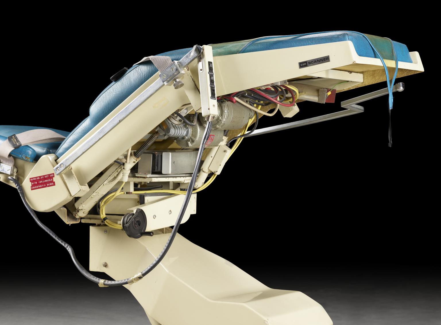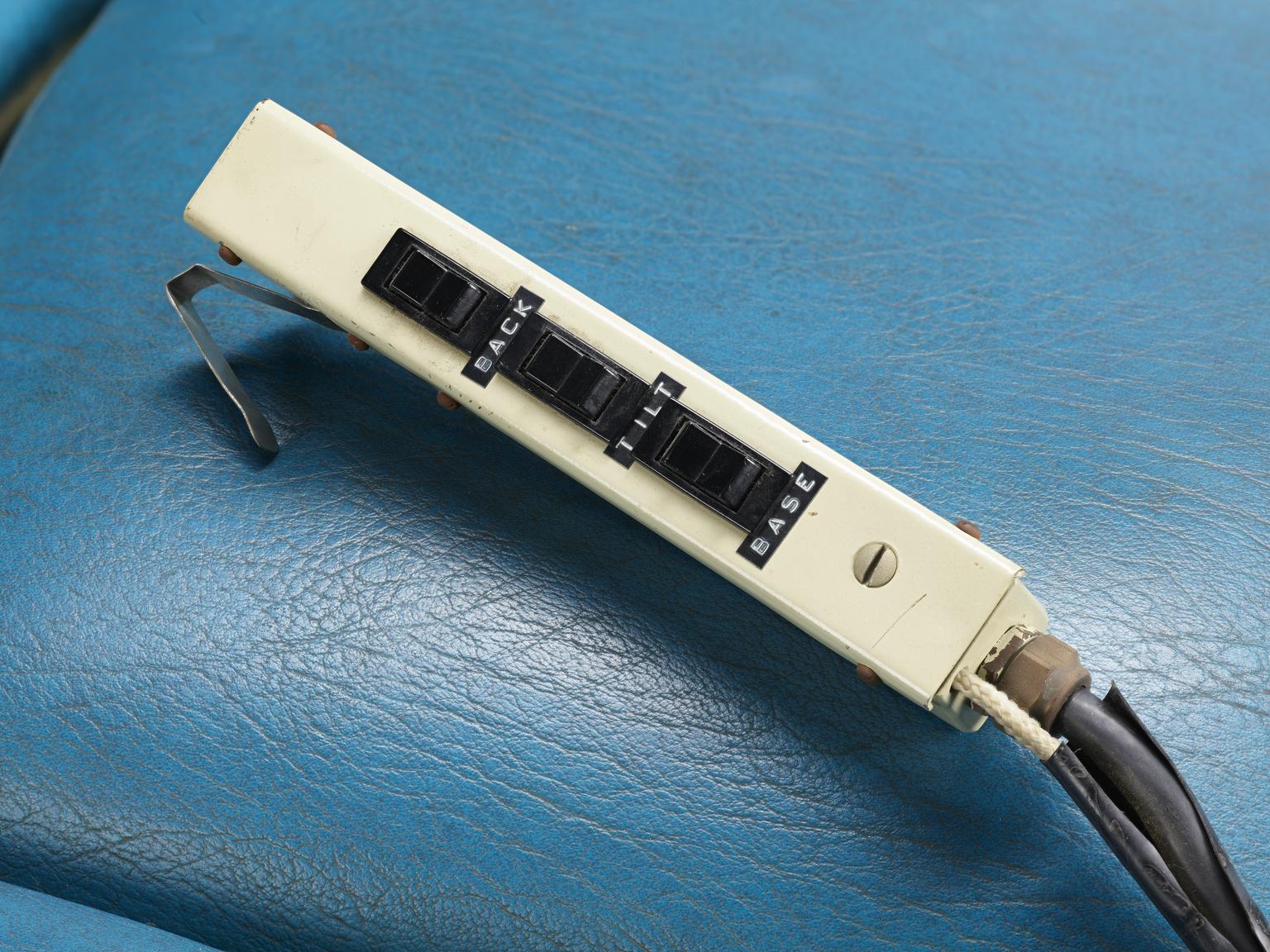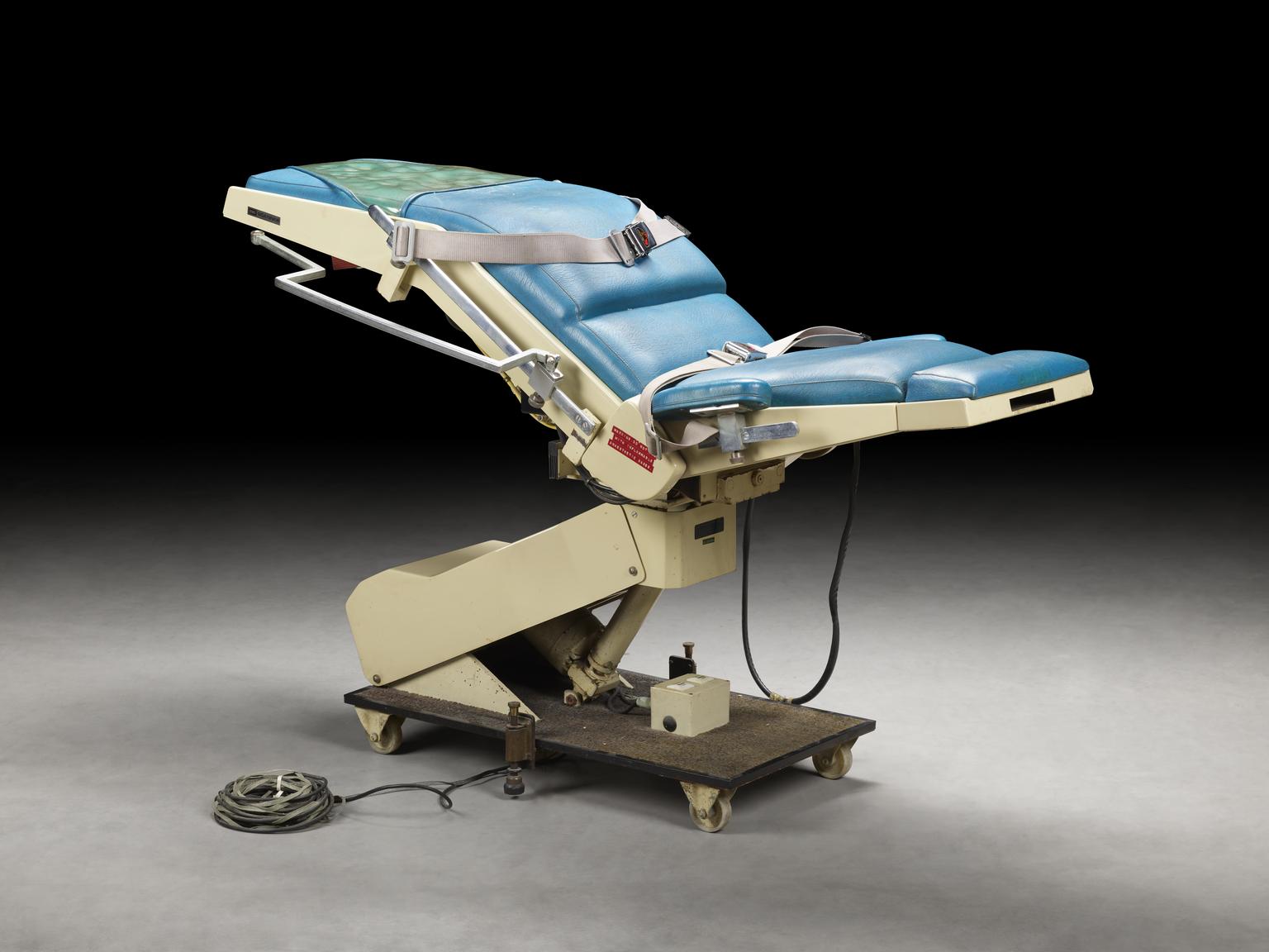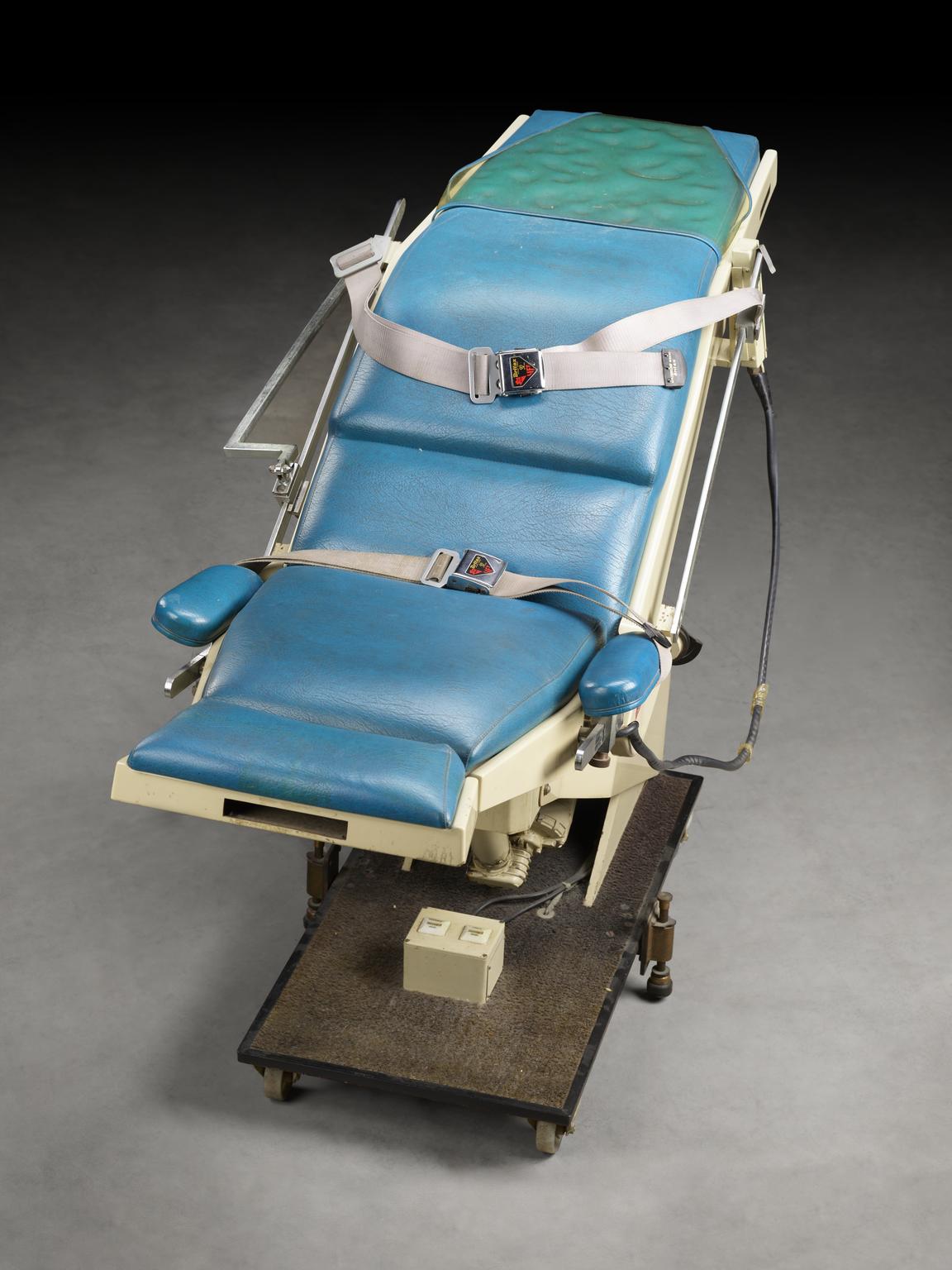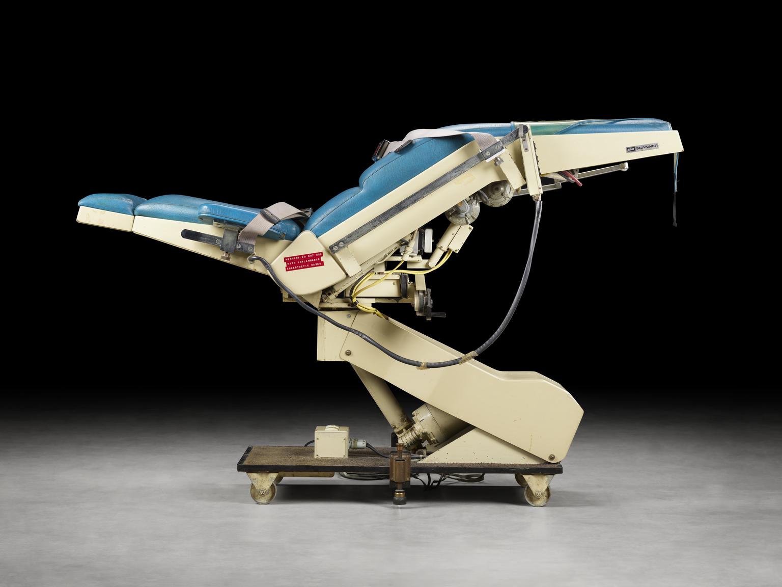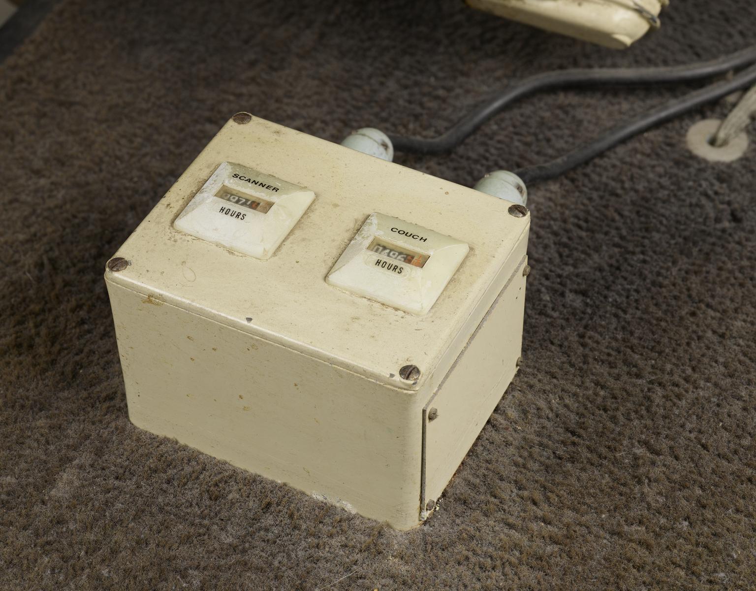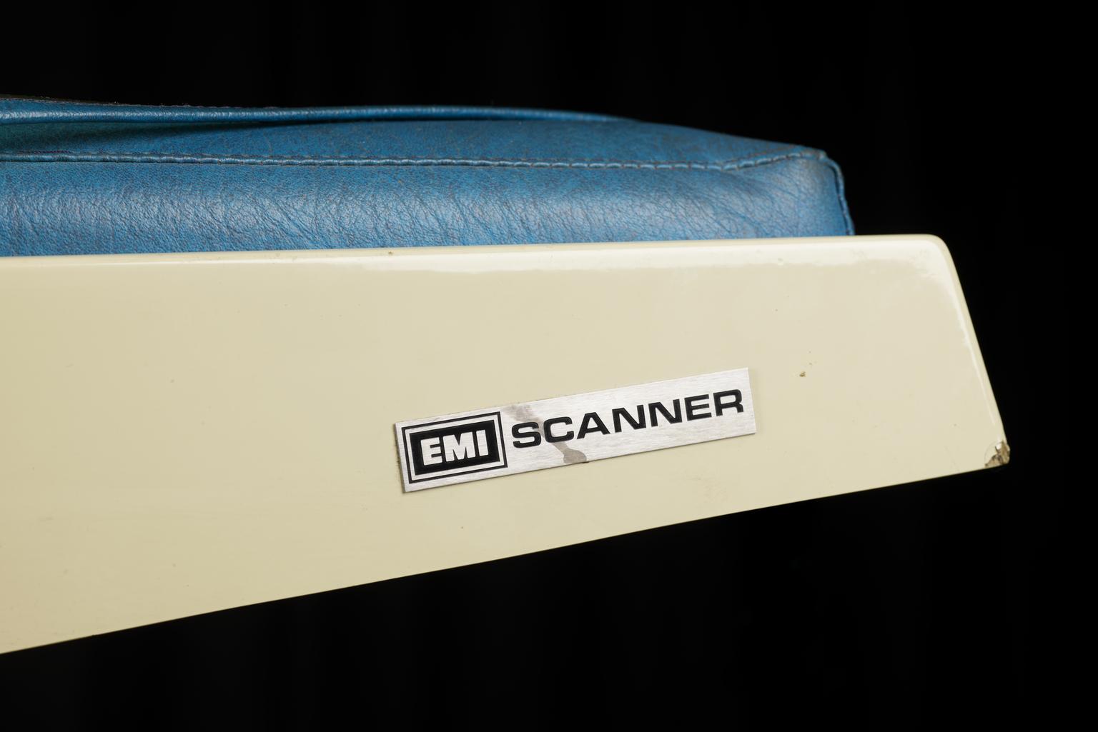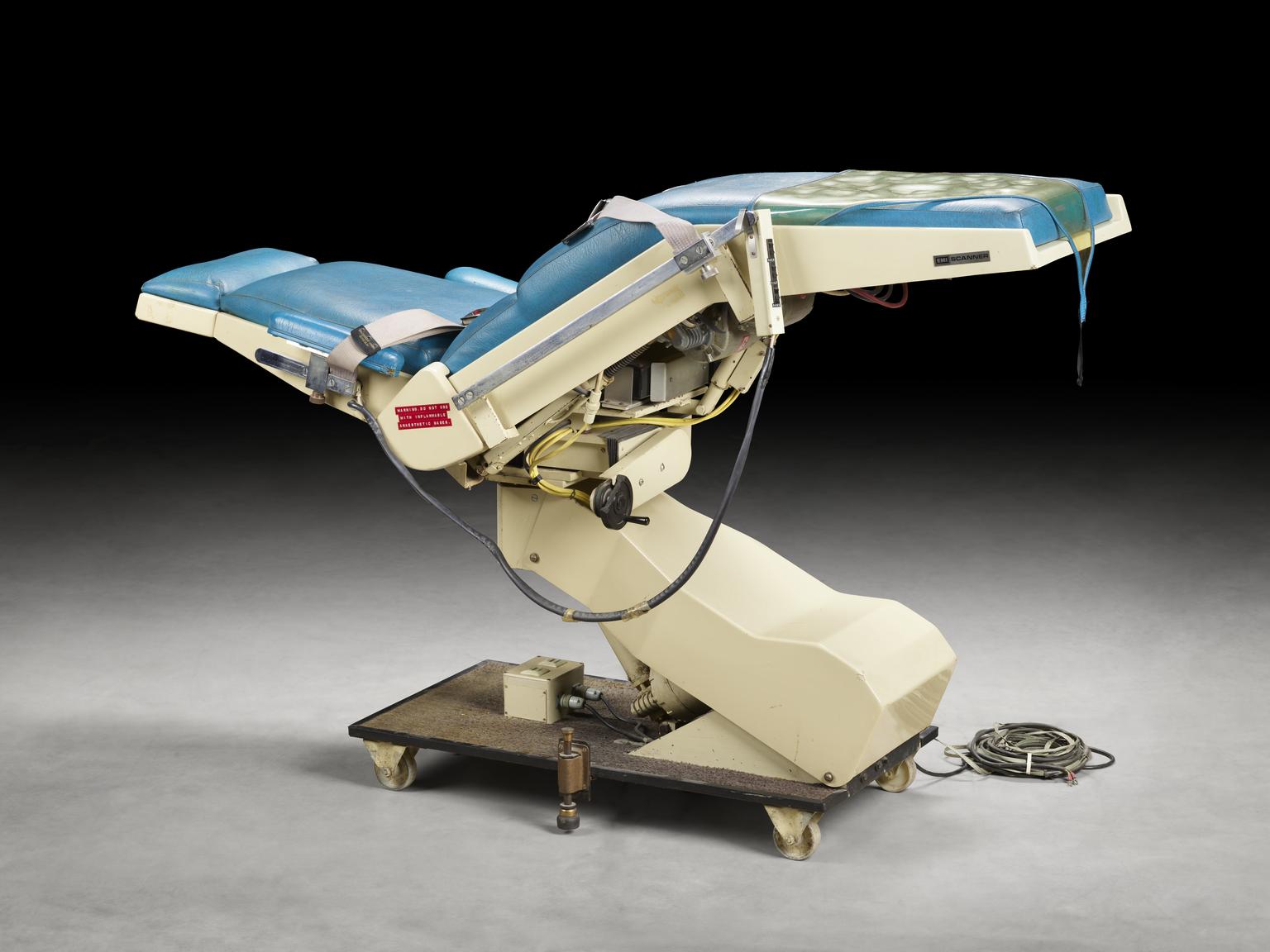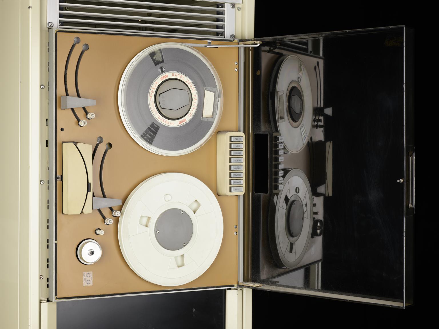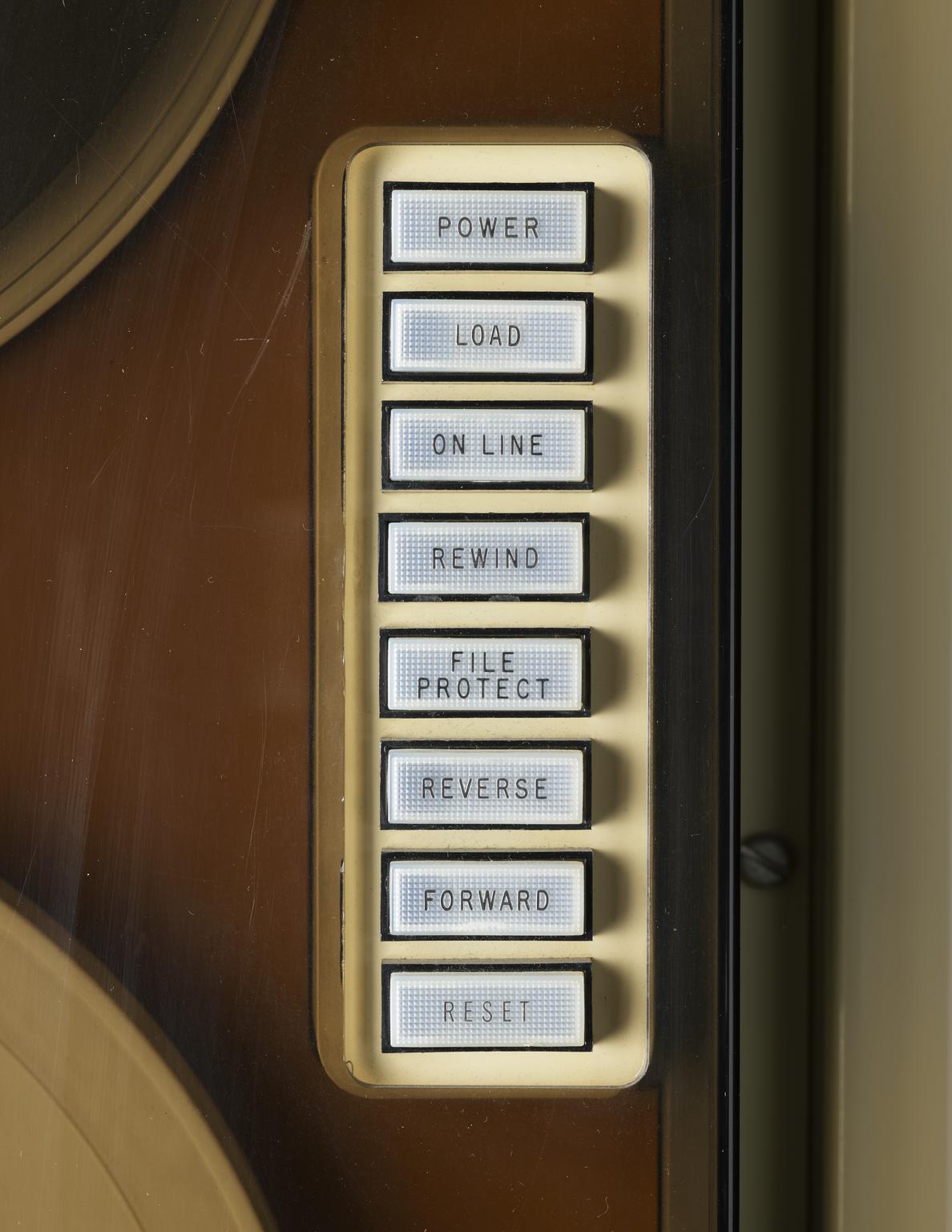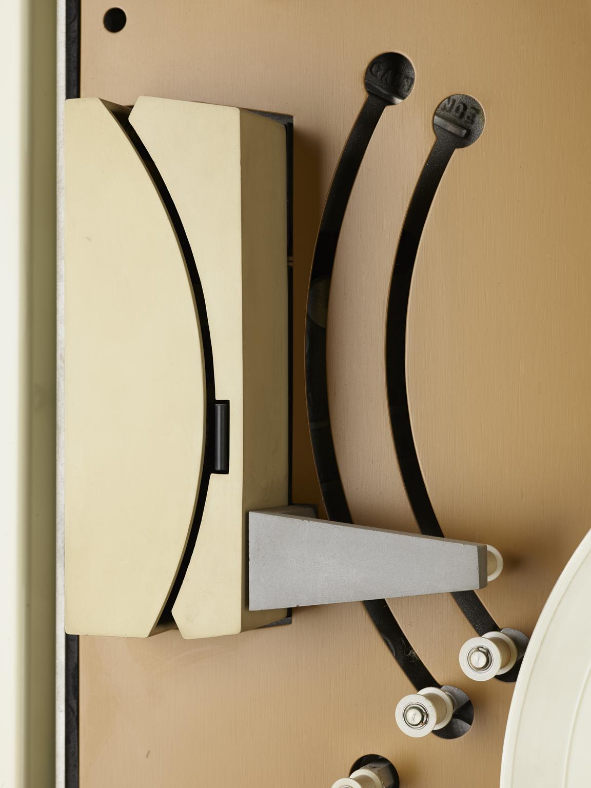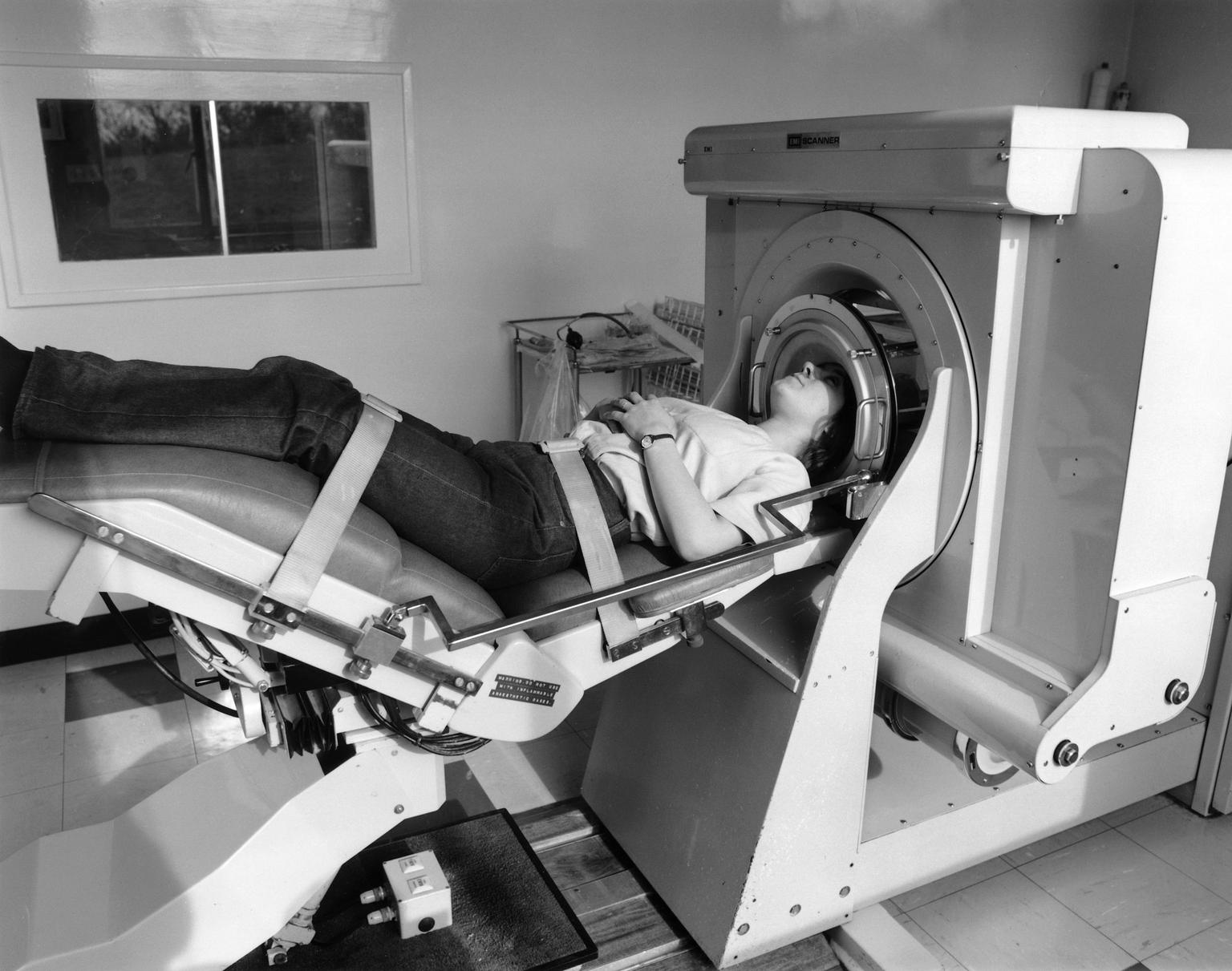
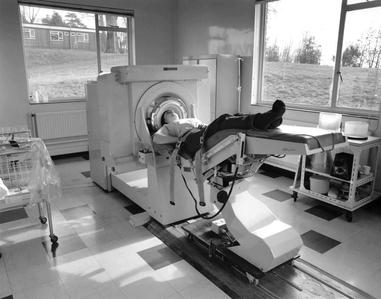
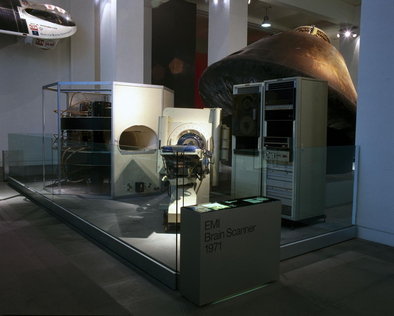
Couch unit for EMI brain scanner
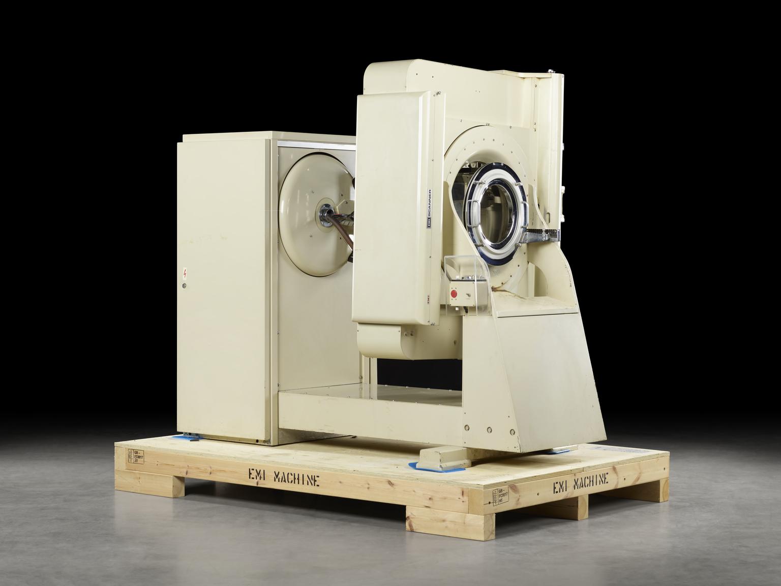
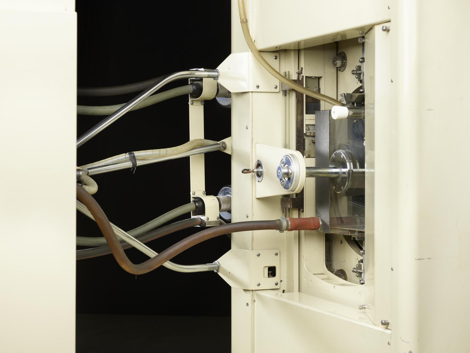
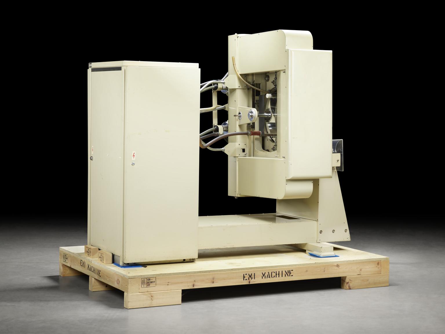
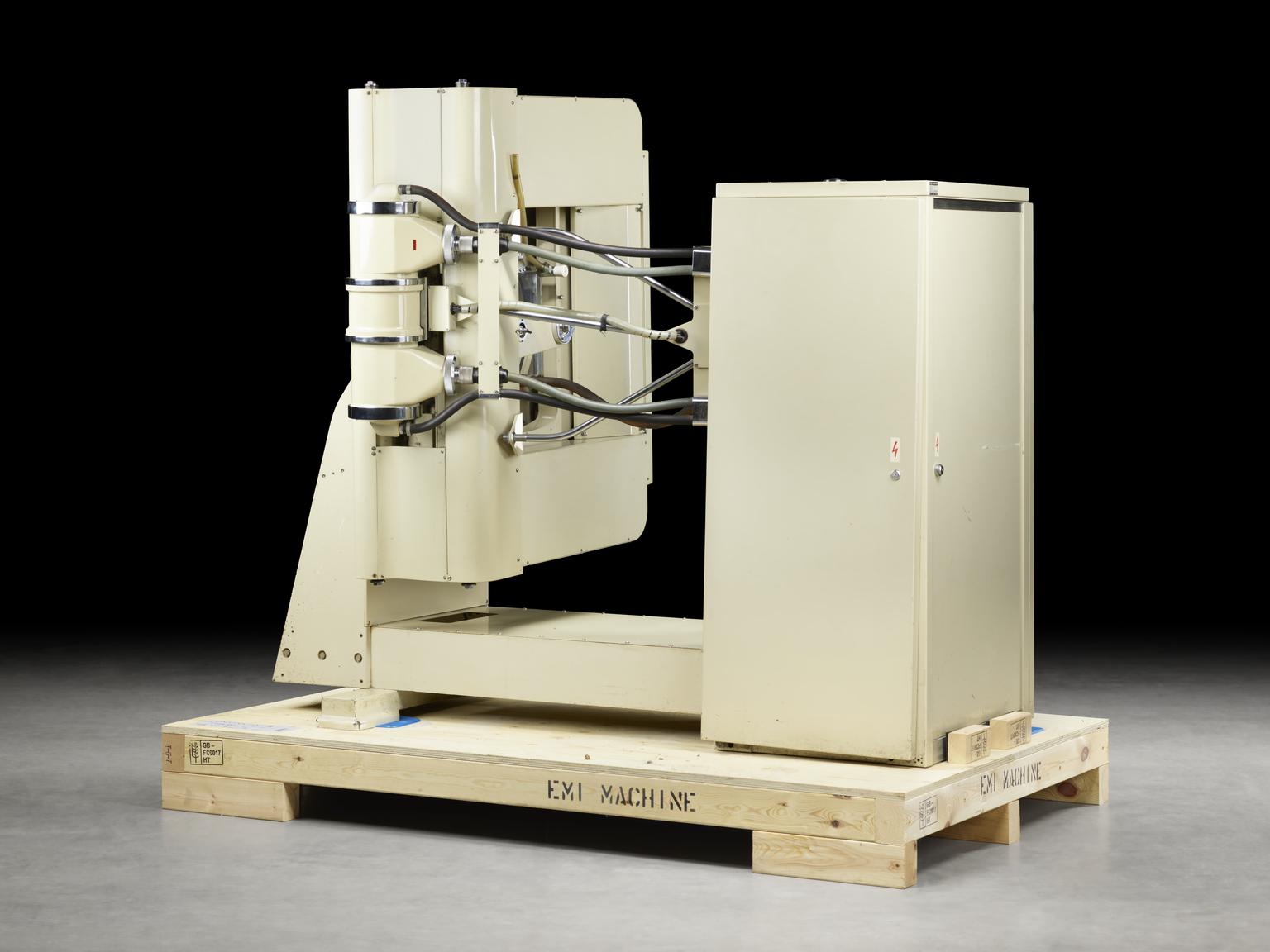
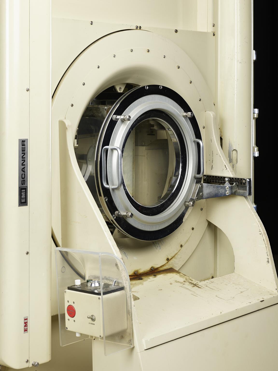
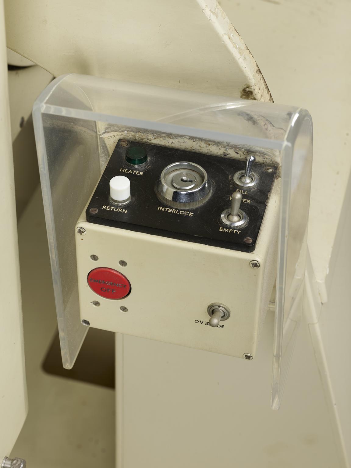
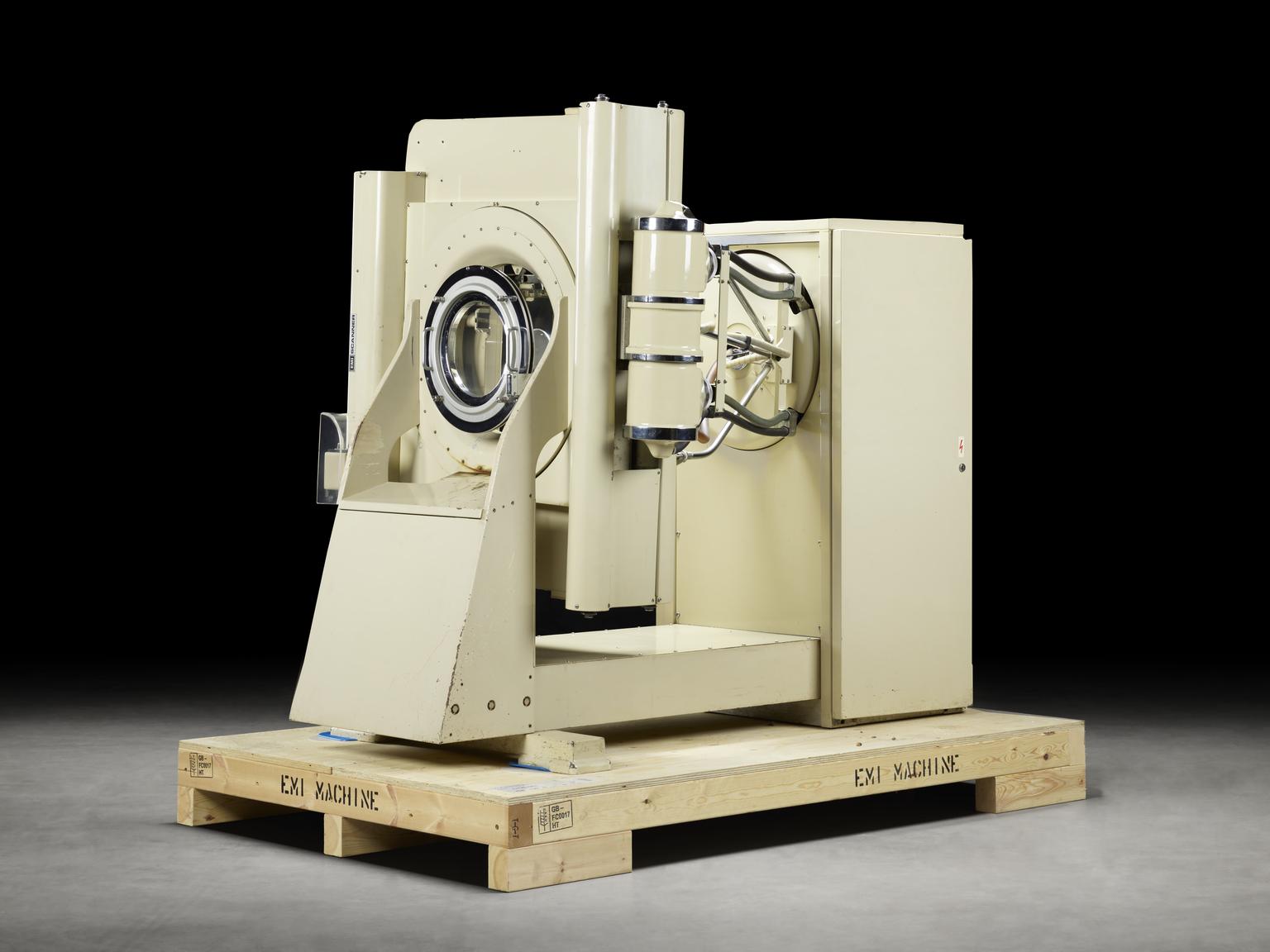
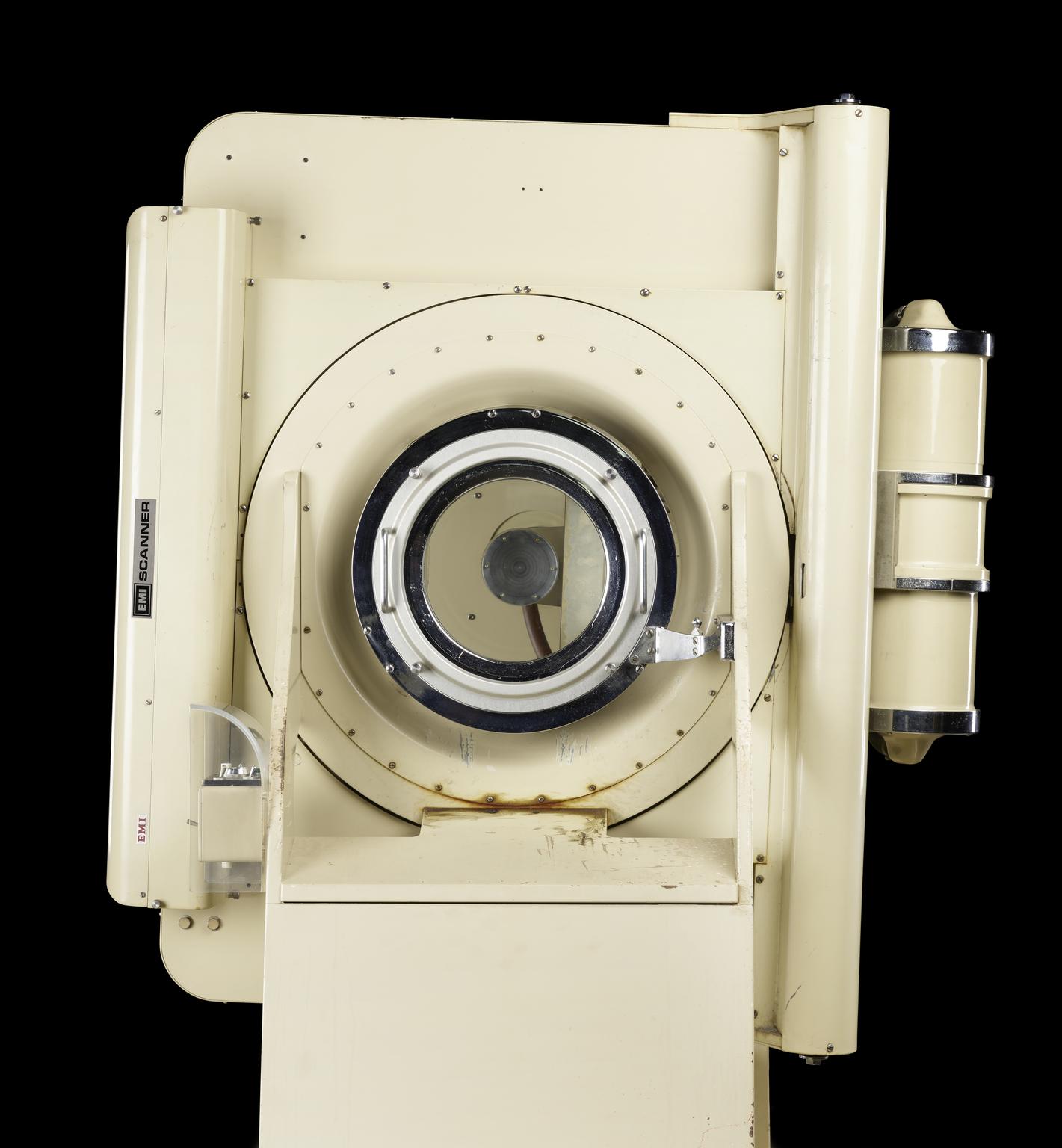
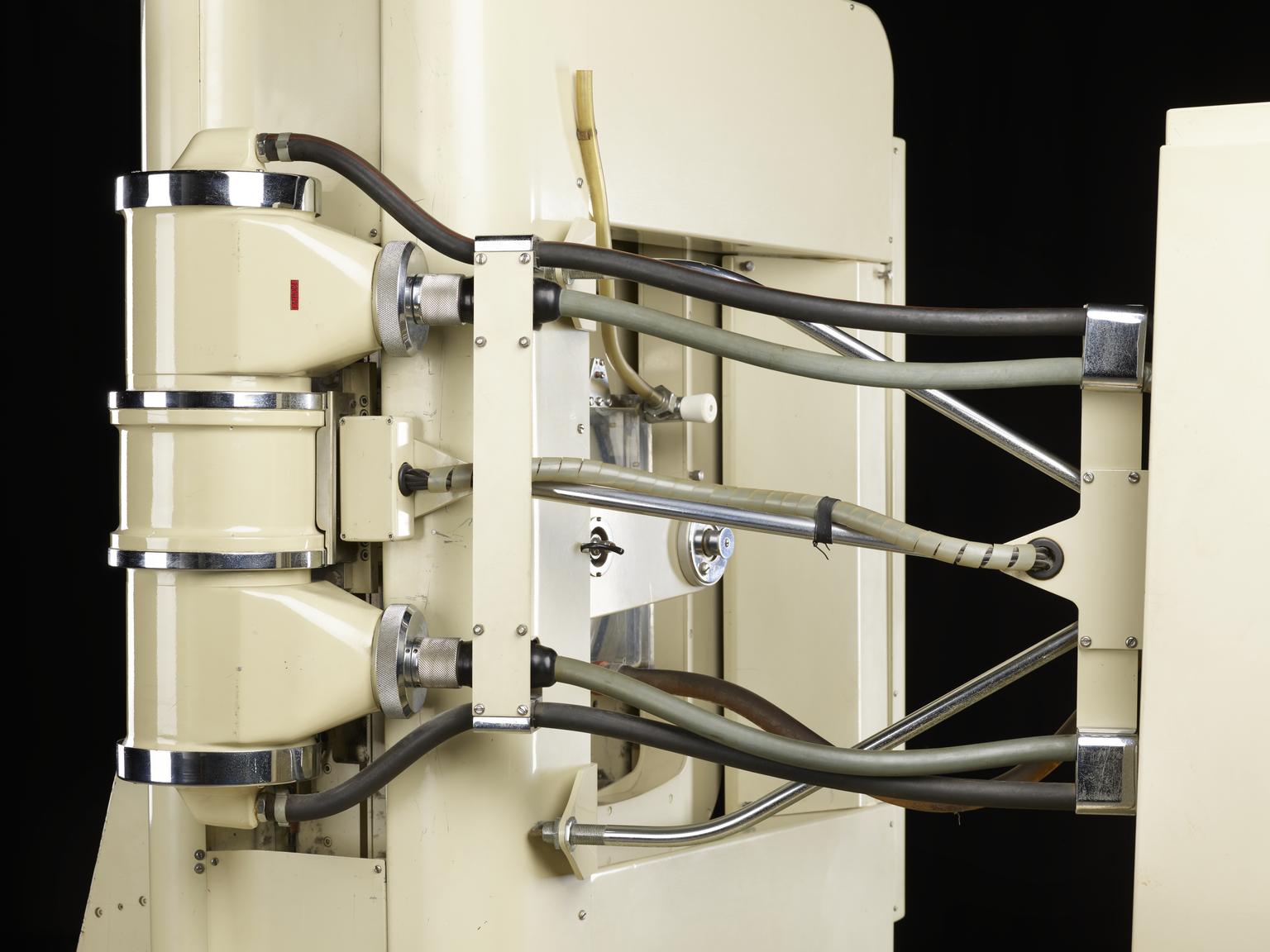
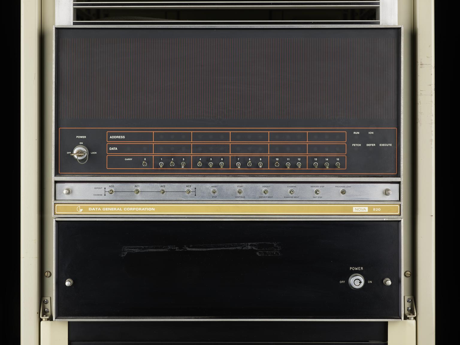
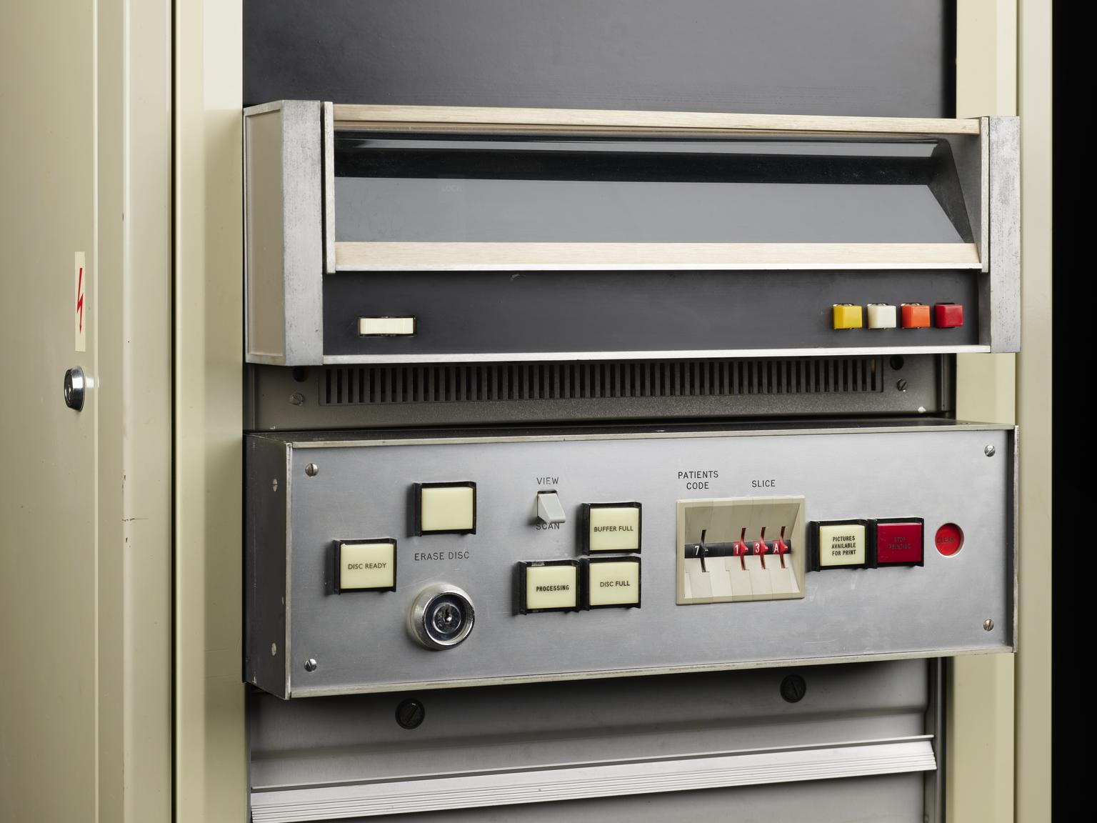
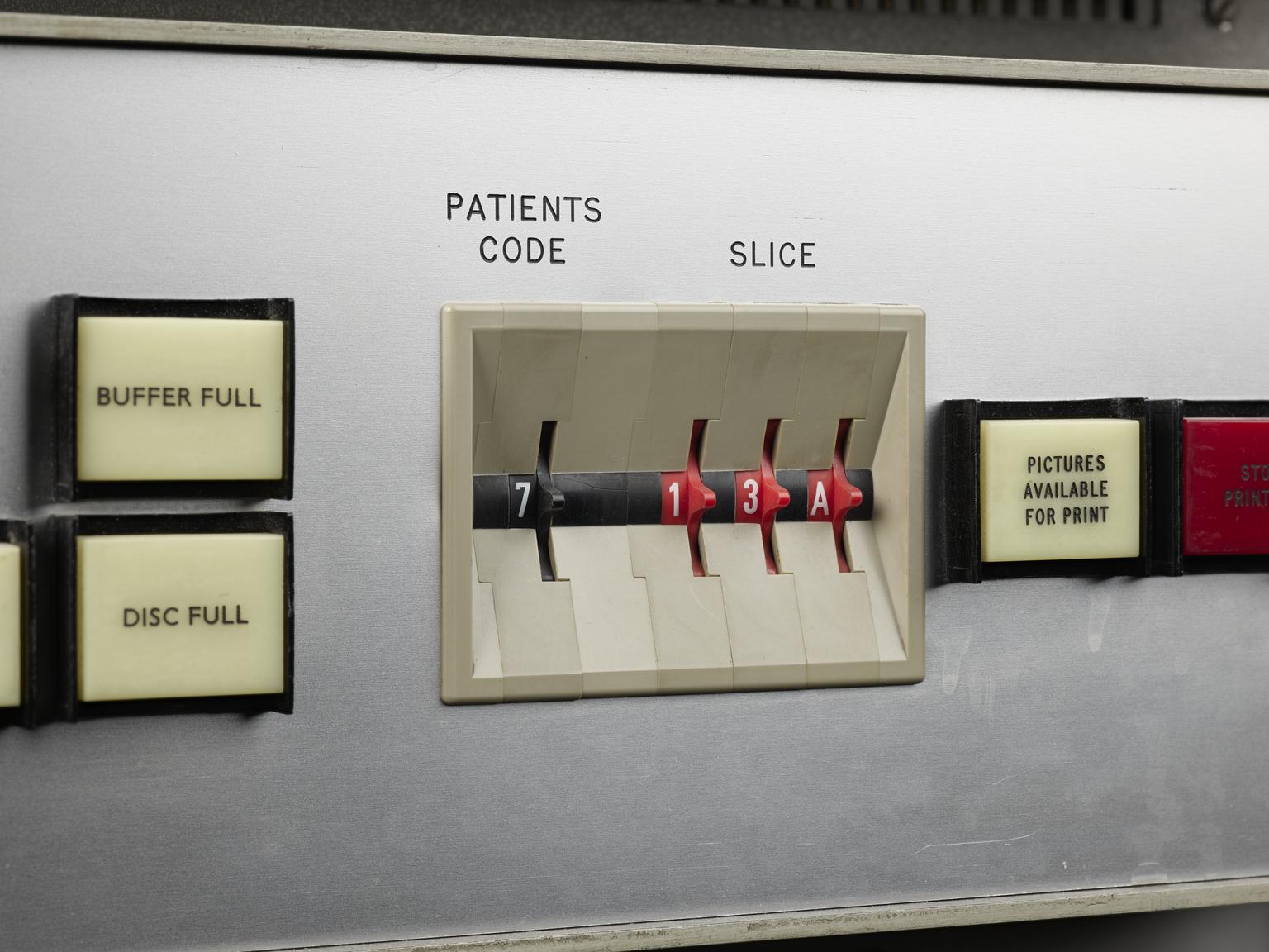
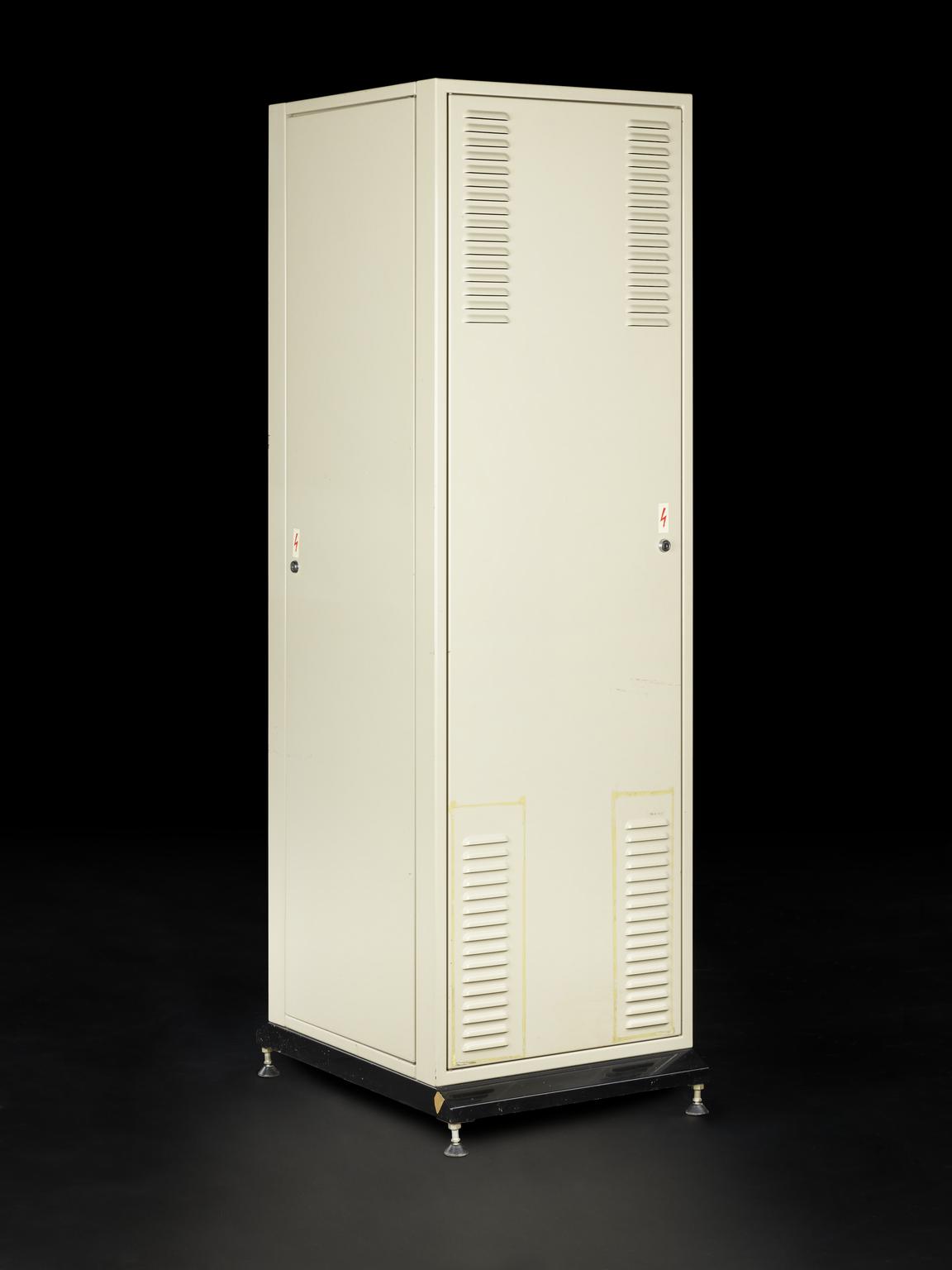
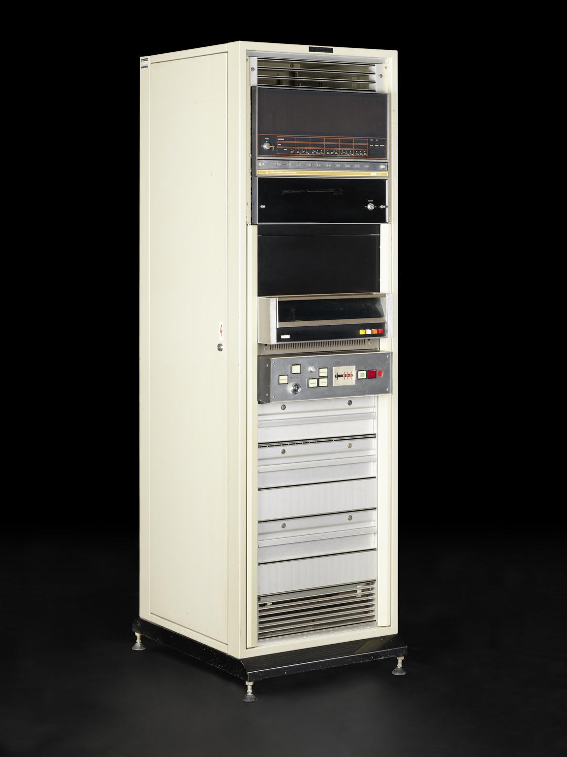
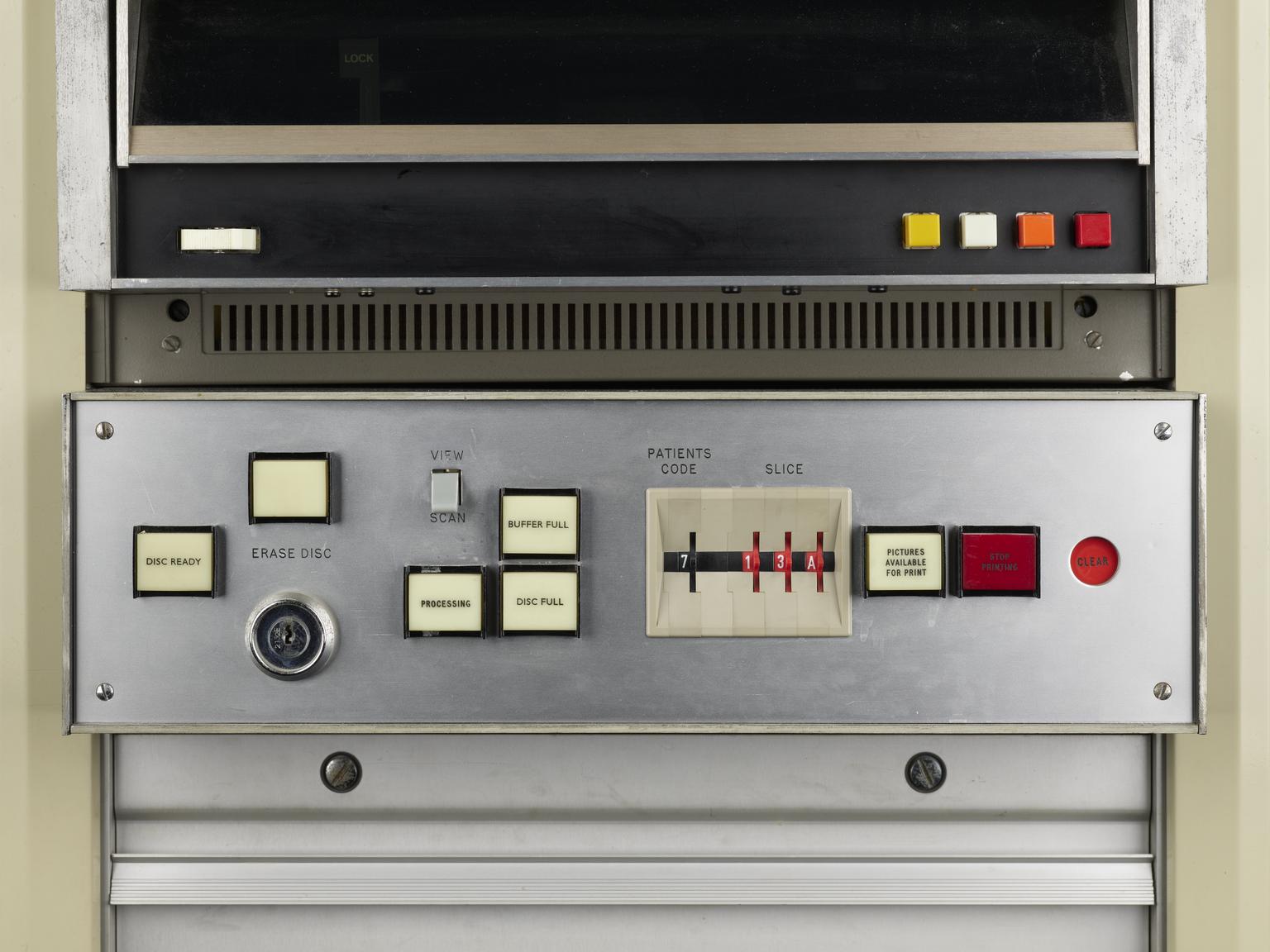
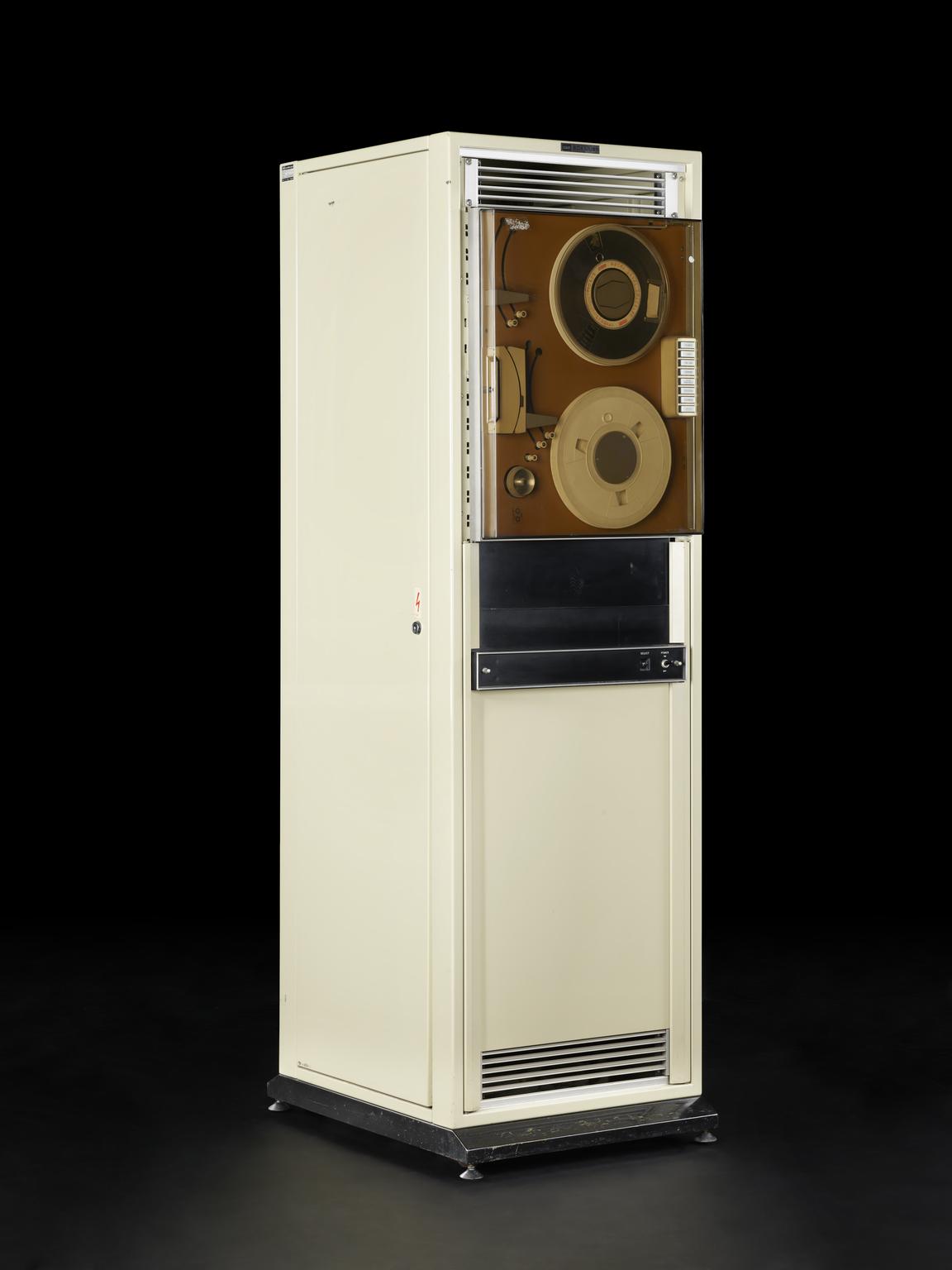
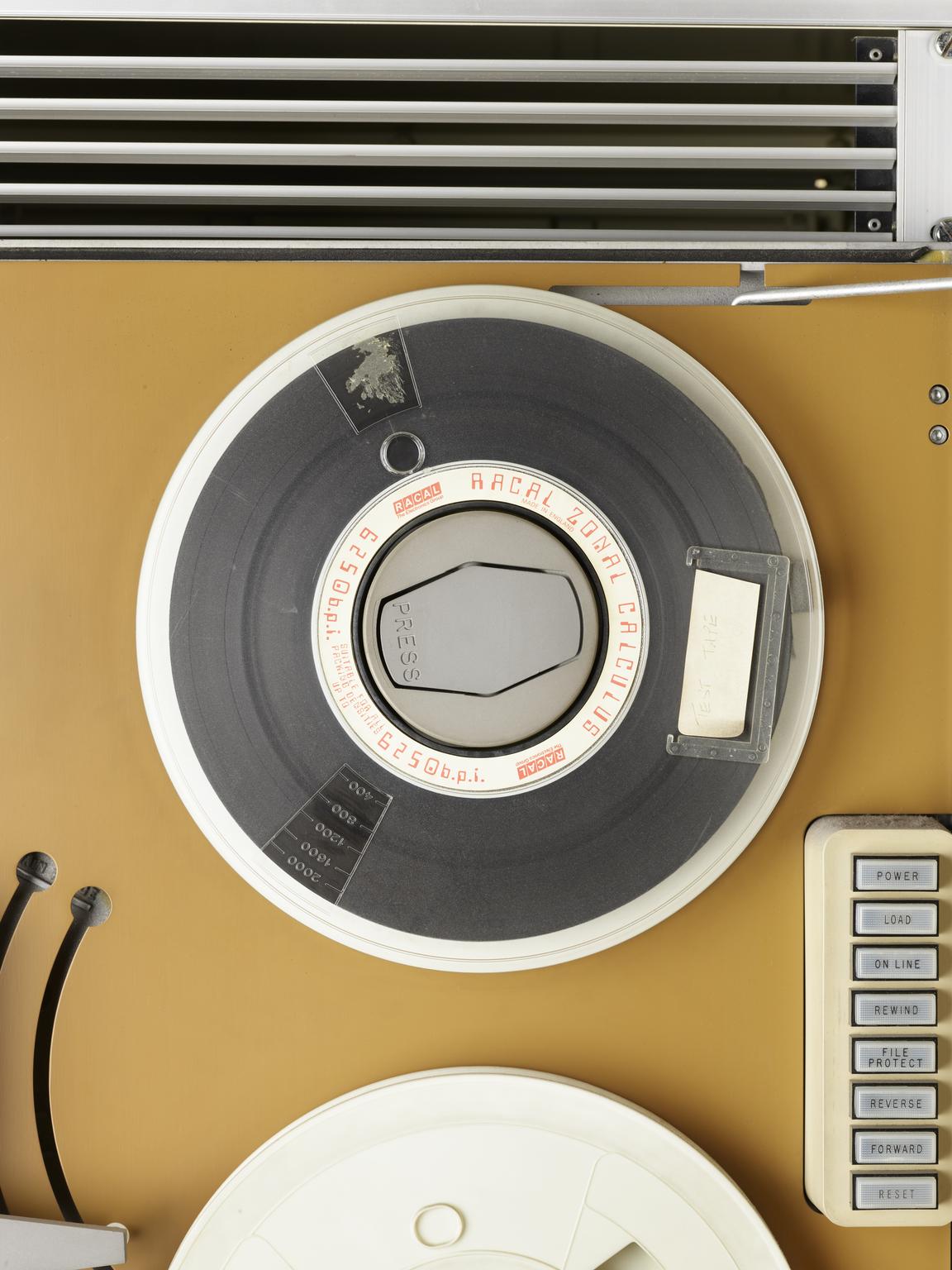
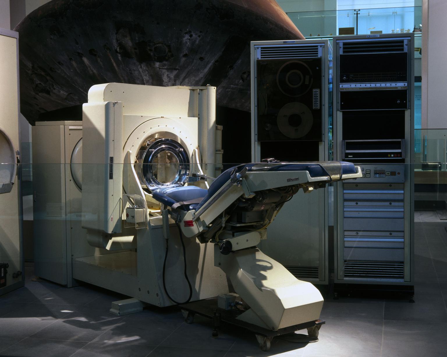
Motorised couch Unit and main gantry for EMI CT brain scanner, installed at the Atkinson Morley's Hospital, Wimbledon in 1971 (the first used clinically), by EMI, Hayes, Middlesex, 1970-1971.
More
Patients would lie on this blue couch unit while the EMI brain scanner produced a computerised tomography (CT) scan of their brain. Developed in 1971 by EMI, detailed pictures of patients’ brains could be seen for the first time. Godfrey Hounsfield (1914-2004) invented the technique, which constructed a picture from measurements made by an X-ray source and detector rotating around the patient. Previously, X-rays could only image the brain after it had received hazardous injections of air or special liquids.
The EMI brain scanner was the first to be adopted in substantial numbers for medicine. Today, Magnetic Resonance Imaging (MRI) has taken over much of the work of CT scanning. This example was installed at Atkinson Morley's Hospital in Wimbledon, London, a specialist neuroscience hospital.
- Materials:
- frame, steel
- Object Number:
- 1980-811 Pt1
- type:
- couch unit
- Image ©
- The Board of Trustees of the Science Museum