Miniature chest x-ray, London, England, 1953-1975

Collection of miniature chest x-rays (6 single frames Collection of miniature chest x-rays (6 single frames

Creative Commons LicenseThis image is released under a CC BY-NC-SA 4.0 Licence

Buy this image as a print 

Buy

License this image for commercial use at Science and Society Picture Library

License

Creative Commons LicenseThis image is released under a CC BY-NC-SA 4.0 Licence

Buy this image as a print 

Buy

License this image for commercial use at Science and Society Picture Library

License

Collection of miniature chest x-rays (6 single frames
Science Museum Group Collection
© The Board of Trustees of the Science Museum

Collection of miniature chest x-rays (6 single frames
Science Museum Group Collection
© The Board of Trustees of the Science Museum

Collection of miniature chest x-rays (6 single frames in paper mounts, 2 rolls of film (35mm & 120 roll film) & 2 paper prints) taken by the North East Thames Mass Radiography Unit, London, 1953-1975

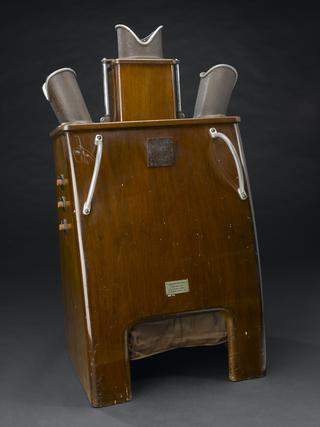
The x-rays produced by miniature radiography are only 100 mm high. The image is viewed on a projector. If the miniature x-ray photograph was inconclusive, the patient would be sent for a full chest X-ray.

Mass miniature radiography was in use in the United Kingdom from 1936 onwards. Doctors using radiography and x-rays found that some of those who appeared ‘healthy’ still showed signs of disease, such as lesions in the lungs caused by pulmonary tuberculosis. Today, x-ray departments are found in hospitals and images are taken of every part of the body instead of just the chest.

Details

Category:
Radiomedicine
Object Number:
1981-2087 Pt1
Materials:
cellulose acetate and cardboard
Measurements:
overall: 100 mm
type:
x-ray photograph

Parts

Miniature chest x-ray, London, England, 1953-1975

Miniature chest x-ray, London, England, 1953-1975

6 single frames in paper mounts, from collection of miniature chest x-rays, taken by the North East Thames Mass Radiography Unit, London, 1953-1975

More

The x-rays produced by miniature radiography are only 100 mm high. The image is viewed on a projector. If the miniature x-ray photograph was inconclusive, the patient would be sent for a full chest X-ray.

Mass miniature radiography was in use in the United Kingdom from 1936 onwards. Doctors using radiography and x-rays found that some of those who appeared ‘healthy’ still showed signs of disease, such as lesions in the lungs caused by pulmonary tuberculosis. Today, x-ray departments are found in hospitals and images are taken of every part of the body instead of just the chest.

Materials:
cellulose acetate and cardboard
Object Number:
1981-2087 Pt1/1
type:
x-ray photograph
Miniature chest x-ray, London, England, 1953-1975

Miniature chest x-ray, London, England, 1953-1975

2 rolls of film, one 35mm & one 120 roll film, from collection of miniature chest x-rays taken by the North East Thames Mass Radiography Unit, London, 1953-1975

More

The x-rays produced by miniature radiography are only 100 mm high. The image is viewed on a projector. If the miniature x-ray photograph was inconclusive, the patient would be sent for a full chest X-ray.

Mass miniature radiography was in use in the United Kingdom from 1936 onwards. Doctors using radiography and x-rays found that some of those who appeared ‘healthy’ still showed signs of disease, such as lesions in the lungs caused by pulmonary tuberculosis. Today, x-ray departments are found in hospitals and images are taken of every part of the body instead of just the chest.

Materials:
cellulose acetate and cardboard
Object Number:
1981-2087 Pt1/2
type:
x-ray photograph
Miniature chest x-ray, London, England, 1953-1975

Miniature chest x-ray, London, England, 1953-1975

2 paper prints, from collection of miniature chest x-rays taken by the North East Thames Mass Radiography Unit, London, 1953-1975

More

The x-rays produced by miniature radiography are only 100 mm high. The image is viewed on a projector. If the miniature x-ray photograph was inconclusive, the patient would be sent for a full chest X-ray.

Mass miniature radiography was in use in the United Kingdom from 1936 onwards. Doctors using radiography and x-rays found that some of those who appeared ‘healthy’ still showed signs of disease, such as lesions in the lungs caused by pulmonary tuberculosis. Today, x-ray departments are found in hospitals and images are taken of every part of the body instead of just the chest.
Materials:
cellulose acetate and cardboard
Object Number:
1981-2087 Pt1/3
type:
x-ray photograph