![]() This image is released under a CC BY-NC-SA 4.0
Licence
This image is released under a CC BY-NC-SA 4.0
Licence
Buy this image as a print
BuyLicense this image for commercial use at Science and Society Picture Library
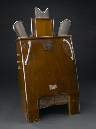
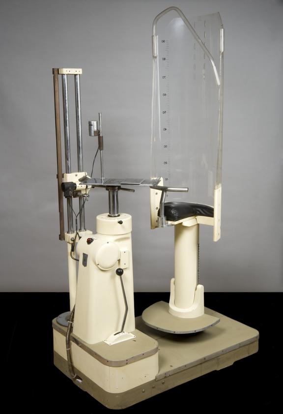
LicenseTransverse Axial Tomographic Unit by Zuder of Turin, 1957
Science Museum Group Collection
© The Board of Trustees of the Science Museum
Transverse Axial Tomographic Unit by Zuder of Turin, 1957
This X-ray unit can locate and then show in 3-D the size and site of a tumour prior to radiotherapy. It was used for cancer research studies at the Holt Radium Institute at the Christie Hospital in Manchester. Transverse axial tomography was a complex process which was restricted to specialist departments such as the Holt Institute. The Institute is still at the forefront of medical research.
Tomography originated in the 1930s. The technique for obtaining radiographs of horizontal sections of the thorax or trunk was patented in 1937 in the UK by William Watson. Italian radiologist Alessandro Vallebona (1899-1987) described a similar technique he called ‘X-ray transverse axial stratigraphy’, also in the 1930s. Vallebona had an X-ray source and a film running in opposite directions around a patient. Only those lying in the plane of rotation were sharply focused. They represented a ‘slice’ of the body.
This machine was made by Zuder of Turin. Tomography was much used as a diagnostic tool until the late 1970s when it was replaced by the Computed Tomography (CT) scanner developed by Godfrey Hounsfield at EMI.
Details
- Category:
- Radiomedicine
- Object Number:
- 1991-431
- Measurements:
-
overall: 2000 mm x 760 mm x 1020 mm,
- credit:
- Christie Hospital and Holt Radium Instit