The main part of the Frank Horrocks collection of microscopical preparations
The main part of the Frank Horrocks collection of microscopical preparations.
The Frank Horrocks collection of microscopical preparations.
The main part of the Frank Horrocks collection of microscopical preparations.
Single pitchblende slide from, The Frank Horrocks collection of microscopical preparations (1979-368/28/20/1).
Wooden cabinet containing 209 microscope slides of histology specimens, prepared by Flatters & Garnett; A. J. Doherty; C. M. Topping; H Vial; A Nield; C. Allott; Wheeler; Watson and sons; Sidwell; Smith, Beck & Beck; C X H Medical School; Rosseter; Penney; H W G; and Husbands, England, 1883-1918
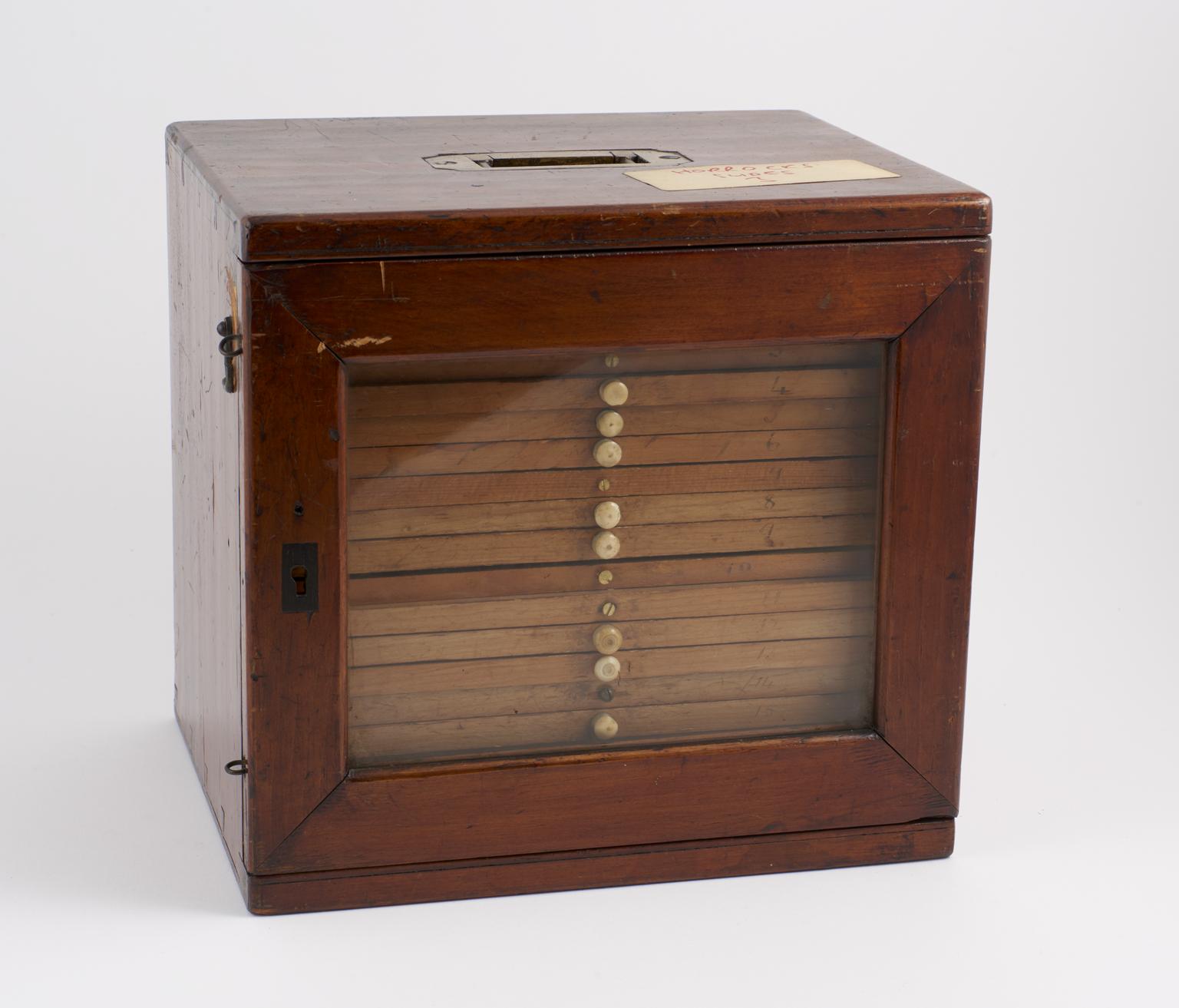
Wooden cabinet containing 203 microscope slides of histology specimens, prepared by Flatters & Garnett; A. J. Doherty; C. M. Topping; H Vial; A Nield; C. Allott; Wheeler; Watson and sons; Sidwell; Smith, Beck & Beck; C X H Medical School; Rosseter; Penney; H W G; and Husbands, England, 1883-1918
Microscope slide containing part of a human ovary, unsigned, England, 1883-1918
Microscope slide containing part of a human umbilical cord, prepared by Flatters & Garnett, England, 1883-1918
Microscope slide containing part of a human pancreas, prepared by Flatters & Garnett, England, 1883-1918
Microscope slide containing human muscle, unsigned, England, 1883-1918
Microscope slide containing a section of the human auriele, unsigned, England, 1883-1918
Microscope slide containing human blood, prepared by Flatters & Garnett, England, 1883-1918
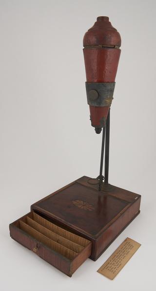
Wooden slide box containing 10 trays with 150 slides of parasites, Human anatomy, histology and pathology, part of the Frank Horrocks slide collection, Middlesex, England, United Kingdom, 1860-1933
Microscope slide containing an unknown Human tissue sample, no labels or markings, unsigned, Middlesex, England, United Kingdom, 1860-1933
Microscope slide containing a sample of a Human Myelin, the label is inscribed: “… Maida Vale Hos. Name Simpkins P. M. Ref. Wallin MYELIN”, unsigned, Middlesex, England, United Kingdom, 1860-1933
Microscope slide containing a sample of a Human blood corpuscles, the label is inscribed: “BLOOD CORPUSCLES (ANAEMIC) 20/2/39”, unsigned, Middlesex, England, United Kingdom, 1939
Microscope slide containing a sample of a Human Aorta, the label is inscribed: “TS. Aorta St. haematoxylin eosin …”, unsigned, Middlesex, England, United Kingdom, 1860-1933
Microscope slide containing a sample of a Gumma from a human liver, the label is inscribed: “Gumma of Liver G. P. I.”, unsigned, Middlesex, England, United Kingdom, 1860-1933
Microscope slide containing a sample of a Human Ovarium dermoid, with two labels; one of which is inscribed: “H. Boecker Ovarium Dermoid Wetzlar.”, and the other label is inscribed: “Mikroskopische Vama Institut”, prepared by H. Boecker, Middlesex, England, United Kingdom, 1860-1933
Microscope slide containing a sample of a Human Lobar Pneumonia Grey Hepatization, with two labels; one of which is inscribed: “PATH Lobar Pneumonia Grey Hepatization”, and the other label is inscribed: “4 Lobar Pneumonia grey hepatization”, unsigned, Middlesex, England, United Kingdom, 1860-1933
Microscope slide containing a sample of Human lymph cells, the label is inscribed: “LUMPHO GRANUOLA LYMPH CELLS. PUS CELLS. MUCOUS. 20/10/40”, unsigned, Middlesex, England, United Kingdom, 1940
Microscope slide containing a sample of a Human Syphilitic Aortitis with two labels; one of which is inscribed: “PATH Syphilitic Aortitis”, and the other label is illegible, unsigned, Middlesex, England, United Kingdom, 1860-1933
Microscope slide containing a sample of Human Tubercular Broncho-Pneumonia, the label is inscribed: “Tubercular Broncho-Pneumonia”, unsigned, Middlesex, England, United Kingdom, 1860-1933
Microscope slide containing an unknown Human tissue sample, glass is marked: “… B 6”, unsigned, Middlesex, England, United Kingdom, 1860-1933
Microscope slide containing a sample of Human tubercle of the hand, the label is inscribed: “20 ‘Sarcoid’ Tubercular of Hand.”, unsigned, Middlesex, England, United Kingdom, 1860-1933
Microscope slide containing a sample of Human tissue, the label is inscribed: “Pathological Laboratory 127 Prot. … carbol fuchsin 16.5.02 University College London”, prepared by University College London, London, England, United Kingdom, 1902
Microscope slide containing a sample of Human E. histolytica iron haemophiliac, the label is inscribed: “… 593 E. histolytica iron Haemophiliac 16.3.26”, unsigned, Middlesex, England, United Kingdom, 1926
Microscope slide containing a sample of a Human Uvula, with two labels the first label is inscribed: “Del Hae. … … ”, the other is inscribed: “382 L. S. Human Uvula”, unsigned, Middlesex, England, United Kingdom, 1860-1933
Microscope slide containing a sample of Human tissue, with two labels the first label is inscribed: “Del Hae. C. Hutchsin”, the other is inscribed: “375”, unsigned, Middlesex, England, United Kingdom, 1860-1933
Microscope slide containing a sample of a Human Stomach Fundus, the label is inscribed: “Medical Research Institute Accra P M 2 “H” Stomach Fundus 25 11.1.33”, prepared by the Medical Research Institute Accra, Ghana, Africa, 1933
Microscope slide containing a sample of a Human Infarct Kidney, the label is inscribed: “PATH Infarct of Kidney”, unsigned, Middlesex, England, United Kingdom, 1860-1933
Microscope slide containing a sample of a Human Mitral Valve, the label is inscribed: “St. Bartholomew’s Hospital Mitral Valve. Malignant Endocarditis”, prepared by St. Bartholomew’s Hospital, London, England, United Kingdom, 1860-1933
Microscope slide containing a sample of a Human B. Abortus, the label is inscribed: “B. Abortus G. Stain 1957”, unsigned, Middlesex, England, United Kingdom, 1957
Microscope slide containing a sample of a Human Kidney, the label is inscribed: “St. Bartholomew’s Hospital LIVER Multipolar Cirrhosis”, prepared by St. Bartholomew’s Hospital, London, England, United Kingdom, 1860-1933. There is a ‘17’ marked on the glass
Microscope slide containing a sample of Carcinoma of the Human liver and stomach, the label is inscribed: “St. Bartholomew’s Hospital Carcinoma of Stomach and Liver”, prepared by St. Bartholomew’s Hospital, London, England, United Kingdom, 1860-1933
Microscope slide containing a sample of a Human Liver bile duct, the label is inscribed: “St. Bartholomew’s Hospital LIVER Bile-duct Carcinoma”, prepared by St. Bartholomew’s Hospital, London, England, United Kingdom, 1860-1933. ‘003’ is marked in pen on the slide
Microscope slide containing a sample of a Human Liver, the label is inscribed: “St. Bartholomew’s Hospital Liver Hodgkin’s Disease”, prepared by St. Bartholomew’s Hospital, London, England, United Kingdom, 1860-1933. ‘260’ is marked in pen on the slide
Microscope slide containing a sample of a Human Liver, the label is inscribed: “St. Bartholomew’s Hospital Liver Hodgkin’s Disease”, prepared by St. Bartholomew’s Hospital, London, England, United Kingdom, 1860-1933. ‘44’ is marked in pen on the slide
Microscope slide containing a sample of a Human Pericarditis, the label is inscribed: “St. Bartholomew’s Hospital Rh. Pericarditis”, prepared by St. Bartholomew’s Hospital, London, England, United Kingdom, 1860-1933. ‘44’ is marked in pen on the slide
Microscope slide containing a sample of a Human Tubercle, the label is inscribed: “St. Bartholomew’s Hospital … Tubercle”, prepared by St. Bartholomew’s Hospital, London, England, United Kingdom, 1860-1933. ‘141’ is marked in pen on the slide
Microscope slide containing a sample of a Human Malignant granular cyst, the label is inscribed: “Clinical Research Association Limited Malignant Granular Cyst Watergate House, Adelphi, W. C”, prepared by the Clinical Research Association, London, England, United Kingdom, 1860-1933
Microscope slide containing a sample of a Human Kidney, the label is inscribed: “St. Bartholomew’s Hospital LIVER Congenital ∑.”, prepared by St. Bartholomew’s Hospital, London, England, United Kingdom, 1860-1933. ‘220’ is marked in pen on the slide
Microscope slide containing a sample of a Human gland, the label is inscribed: “St. Bartholomew’s Hospital Gland Hodgkin’s Disease.”, prepared by St. Bartholomew’s Hospital, London, England, United Kingdom, 1860-1933. ‘460’ is marked in pen on the slide
Microscope slide containing a sample of Human Rheumatic Myocarditis, the label is inscribed: “St. Bartholomew’s Hospital Rheumatic Myocarditis”, prepared by St. Bartholomew’s Hospital, London, England, United Kingdom, 1860-1933. ‘222’ is marked in pen on the slide
Microscope slide containing a sample of Human Pneumococcal pericarditis, the label is inscribed: “St. Bartholomew’s Hospital Pneumococcal Pericarditis”, prepared by St. Bartholomew’s Hospital, London, England, United Kingdom, 1860-1933. ‘170’ is marked in pen on the slide
Microscope slide containing a sample of Human Syphilitic Myocarditis Gumma, the label is inscribed: “St. Bartholomew’s Hospital Syphilitic Myocarditis Gumma”, prepared by St. Bartholomew’s Hospital, London, England, United Kingdom, 1860-1933
Microscope slide containing a sample of a Human Lymph Node Tubercle, the label is inscribed: “St. Bartholomew’s Hospital Lymph Node Tubercle.”, prepared by St. Bartholomew’s Hospital, London, England, United Kingdom, 1860-1933
Microscope slide containing a sample of a Human tissue, the label is inscribed: “… … … 9 … J. Ross”, prepared by J. Ross, Middlesex, England, United Kingdom, 1860-1933. Writing on label is illegible
Microscope slide containing a sample of Cirrhosis of the Human Liver, with two labels, the first label is inscribed: “G 228”, and the other one is inscribed: “Cirrhosis of Liver”, unsigned, Middlesex, England, United Kingdom, 1860-1933
Microscope slide containing a sample of a Human Membrane, the label is inscribed: “Membrane showing …”, unsigned, Middlesex, England, United Kingdom, 1860-1933
Microscope slide containing a sample of a Human Liver, with two labels, the first label is inscribed: “Liver …”, and the other one is inscribed: “G 229”, unsigned, Middlesex, England, United Kingdom, 1860-1933
Microscope slide containing a sample of a Human skin, with two labels, the first label is inscribed: “195”, and the other one is inscribed: “Object No. 2 Skin Tumours of phasynst”, unsigned, Middlesex, England, United Kingdom, 1860-1933
Microscope slide containing a sample of a Human tissue, with two labels, the first label is inscribed: “… … stained CB”, and the other one is inscribed: “192”, unsigned, Middlesex, England, United Kingdom, 1860-1933
Microscope slide containing a sample of a Human tissue with Lupus, with two labels, the first label is inscribed: “G177”, and the other one is inscribed: “Lupus”, unsigned, Middlesex, England, United Kingdom, 1860-1933
Microscope slide containing a sample of a Human tissue with Typhoid, with two labels, the first label is inscribed: “G164”, and the other one is inscribed: “Typhoid … 10 days”, unsigned, Middlesex, England, United Kingdom, 1860-1933
Microscope slide containing a sample of a Human Gastric Cellulitis, with two labels, the first label is inscribed: “G202”, and the other one is inscribed: “Gastric Cellulitis”, unsigned, Middlesex, England, United Kingdom, 1860-1933
Microscope slide containing a sample of a Human Fibroma of palate, with two labels, the first label is inscribed: “Fibroma of Palate Stained”, and the other one is inscribed: “G162”, unsigned, Middlesex, England, United Kingdom, 1860-1933
Microscope slide containing a sample of a Human Tissue, with two labels, the first label is inscribed: “…”, and the other one is inscribed: “G218”, unsigned, Middlesex, England, United Kingdom, 1860-1933
Microscope slide containing a sample of a Human Liver, with two labels, the first label is inscribed: “Liver Advanced Nutmeg”, and the other one is inscribed: “W. S. H. C .B. P. C. … 18.1.12”, unsigned, Middlesex, England, United Kingdom, 1912
Microscope slide containing a sample of a Human Lung, the label is inscribed: “PATH Infarct of Lung.”, unsigned, Middlesex, England, United Kingdom, 1860-1933
Microscope slide containing a sample of a Human tissue, the label is inscribed: “J. E. McCartney Rib Osteitis deformans”, prepared by J. E. McCartney, Middlesex, England, United Kingdom, 1860-1933
Microscope slide containing a sample of a Human tissue, the label is inscribed: “Anatomy Dept. Loan Slide M. H. M. S”, prepared by Anatomy Dept. Loan Slide M. H. M. S, Middlesex, England, United Kingdom, 1860-1933
Microscope slide containing a sample of a Human Lung, the label is inscribed: “Lung.”, unsigned, Middlesex, England, United Kingdom, 1860-1933
Microscope slide containing a sample of a Human Lympho, the label is inscribed: “Lympho … S. M. Copeman.”, unsigned, Middlesex, England, United Kingdom, 1860-1933
Microscope slide containing a sample of a Human Epiglottis, the label is inscribed: “Section of Epiglottis (…) 1897 no. 1”, unsigned, Middlesex, England, United Kingdom, 1897
Wooden slide box containing 12 trays with 167 slides of various dental histological specimens including Human, part of the Frank Horrocks slide collection, Middlesex, England, United Kingdom, 1934-1939
Wooden cabinet containing microscope slides of histology specimens, prepared by Flatters & Garnett; A. J. Doherty; C. M. Topping; H Vial; A Nield; C. Allott; Wheeler; Watson and sons; Sidwell; Smith, Beck & Beck; C X H Medical School; Rosseter; Penney; H W G; and Husbands, England, 1883-1918