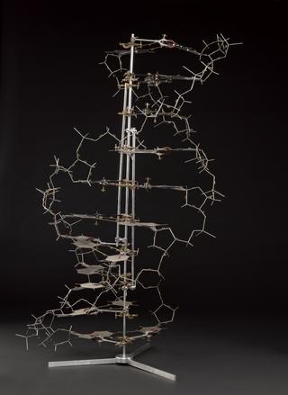
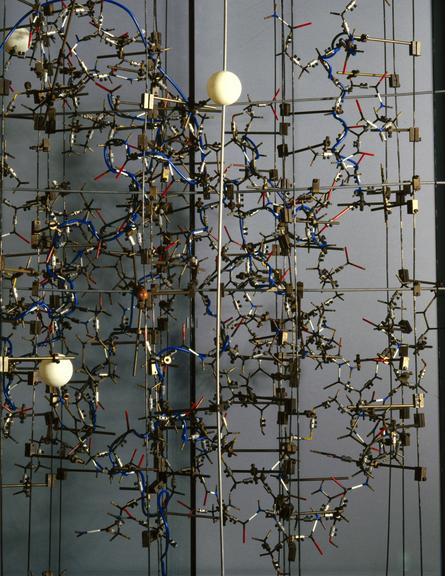
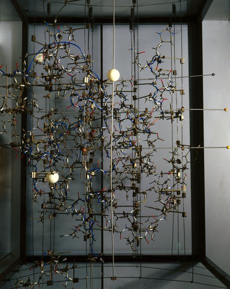
Molecular model showing the structure of insulin


![]() This image is released under a CC BY-NC-SA 4.0
Licence
This image is released under a CC BY-NC-SA 4.0
Licence
License this image for commercial use at Science and Society Picture Library
License![]() This image is released under a CC BY-NC-SA 4.0
Licence
This image is released under a CC BY-NC-SA 4.0
Licence
License this image for commercial use at Science and Society Picture Library
License![]() This image is released under a CC BY-NC-SA 4.0
Licence
This image is released under a CC BY-NC-SA 4.0
Licence
License this image for commercial use at Science and Society Picture Library
License
Science Museum Group Collection
© The Board of Trustees of the Science Museum
Science Museum Group Collection
© The Board of Trustees of the Science Museum
Science Museum Group Collection
© The Board of Trustees of the Science Museum
Model, one of two, made by Dorothy M. Crowfoot Hodgkin c.1967, to show the structure of 2 zinc pig insulin crystals at a resolution of 2.8A
In 1935, Dorothy M Crowfoot Hodgkin (1910-94), a British chemist and crystallographer, published the first X-ray photograph of insulin. Insulin is produced by the pancreas to break down sugars in the body. But Hodgkin and her team were unable to determine the 3-D structure of insulin until 1969, when this model was made. The larger metal balls in the model represent zinc, which was introduced chemically into the protein to decode the rest.
Details
- Category:
- Biochemistry
- Object Number:
- 1991-286/1
- Materials:
- copper (alloy), plastic (unidentified) and steel (metal)
- Measurements:
-
overall: 1000 mm x 780 mm x 930 mm,
- type:
- molecular model
- credit:
- Prof. Dorothy Crowfoot Hodgkin
Related Objects