Tabula IX

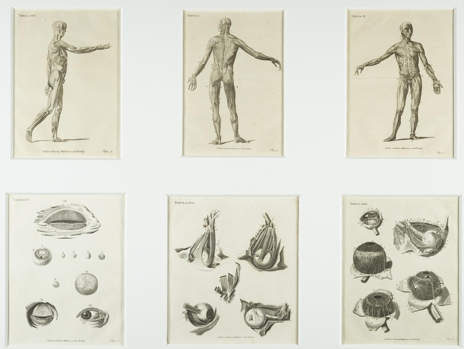
Tabula IX, Tabula X, Tabula XVII, Tabula XLII, Tabula XLIII, Tabula XLIV, Tabula XLV, Tabula XLVI and pages 259-264, from the anatomical treatise 'Schola et Historia Medicinae', by William Rowley, England, edition by Royce, England, 1851-1860

Details

Category:
Art
Object Number:
1980-523/1
type:
prints

Parts

Tabula IX illustrating a front view of the muscles in the human body (male muscular structure)

Tabula IX illustrating a front view of the muscles in the human body (male muscular structure)

Tabula IX illustrating a front view of the muscles in the human body (male muscular structure), taken from the anatomical treatise 'Schola et Historia Medicinae', by William Rowley, England, edition by Royce, England, 1851-1860

More

Showing the muscles of the human body, this print was taken from an anatomical treatise called 'Schola Medicinæ Universalis Nova' or the 'New Universal History and School of Medicine' by William Rowley (1742-1806), an English male midwife, surgeon and anatomist. First published in 1793, the work contained 68 copper engravings of the human body. The work was probably used by medical students studying anatomy. Anatomical prints were useful tools as specific features of the body could be enlarged and picked out, making the structures easier to understand.
Materials:
paper
Object Number:
1980-523/1/1
type:
print
Image ©
The Board of Trustees of the Science Museum
Tabula X illustrating a rear view of muscles in the human body (male muscular structure)

Tabula X illustrating a rear view of muscles in the human body (male muscular structure)

Tabula X illustrating a rear view of muscles in the human body (male muscular structure), taken from the anatomical treatise 'Schola et Historia Medicinae', by William Rowley, England, edition by Royce, England, 1851-1860

More

Showing the muscles of the back of the human body, this print was taken from an anatomical treatise called 'Schola Medicinæ Universalis Nova' or the 'New Universal History and School of Medicine' by William Rowley (1742-1806), an English male midwife, surgeon and anatomist. First published in 1793, the work contained 68 copper engravings of the internal structures of the human body. Anatomical prints were useful tools for the medical students as specific features of the body could be enlarged and picked out, making the structures easier to understand.
Materials:
paper
Object Number:
1980-523/1/2
type:
print
Tabula XVII illustrating a side view muscles in the human body (male muscular structure)

Tabula XVII illustrating a side view muscles in the human body (male muscular structure)

Tabula XVII illustrating a side view muscles in the human body (male muscular structure), taken from the anatomical treatise 'Schola et Historia Medicinae', by William Rowley, England, edition by Royce, England, 1851-1860

More

Showing a side view of the muscles in the human body, this print was taken from an anatomical treatise called 'Schola Medicinæ Universalis Nova' or the 'New Universal History and School of Medicine' by William Rowley (1742-1806), an English male midwife, surgeon and anatomist. A key is used to identify each part of the body. First published in 1793, the work contained 68 copper engravings of the human body. Anatomical prints were useful tools for medical students as specific features of the body could be enlarged and picked out, making the structures easier to understand.
Materials:
paper
Object Number:
1980-523/1/3
type:
print
Tabula XLII illustrating the muscles of the human eye

Tabula XLII illustrating the muscles of the human eye

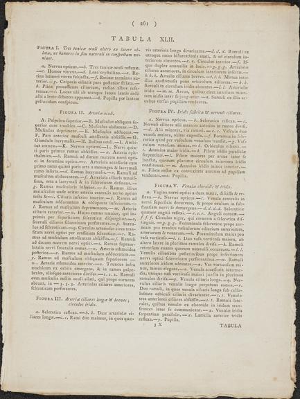
Tabula XLII illustrating the muscles of the human eye, plus pages 259-260, taken from the anatomical treatise 'Schola et Historia Medicinae', by William Rowley, England, edition by Royce, England, 1851-1860

More

Each diagram in the print unpeels a layer of the human eye accompanied by letters used with a key to identify each part. This print was taken from an anatomical treatise called 'Schola Medicinæ Universalis Nova' or the 'New Universal History and School of Medicine' by William Rowley (1742-1806), an English male midwife, surgeon and anatomist. First published in 1793, the work contained 68 copper engravings of the human body. Anatomical prints were useful tools for medical students as specific features of the body could be enlarged and picked out, making the structures easier to understand.

Object Number:
1980-523/1/4
type:
print

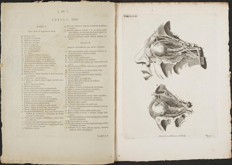
Tabula XLIII illustrating sections through the human eye and optic nerve

Tabula XLIII illustrating cross-sections of the human eye and optic nerve, plus pages 261-262 of the anatomical treatise 'Schola et Historia Medicinae', by William Rowley, England, edition by Royce, England, 1851-1860

More

The structure of the human eye has been revealed in this print, which shows a dissected human face, accompanied by letters used with a key to identify each part. This print was taken from an anatomical treatise called 'Schola Medicinæ Universalis Nova' or the 'New Universal History and School of Medicine' by William Rowley (1742-1806), an English male midwife, surgeon and anatomist. First published in 1793, the work contained 68 copper engravings of the human body. Anatomical prints were useful tools for medical students as specific features of the body could be enlarged and picked out, making the structures easier to understand.
Object Number:
1980-523/1/5
type:
print
Image ©
The Board of Trustees of the Science Museum
Tabula XLIV illustrating the human eye

Tabula XLIV illustrating the human eye

Tabula XLIV illustrating the human eye, eyelid and lens, taken from the anatomical treatise 'Schola et Historia Medicinae', by William Rowley, England, edition by Royce, England, 1851-1860

More

Each part of the human eye, eyelid and lens has been dissected and enlarged in this print. This print was taken from an anatomical treatise called 'Schola Medicinæ Universalis Nova' or the 'New Universal History and School of Medicine' by William Rowley (1742-1806), an English male midwife, surgeon and anatomist. First published in 1793, the work contained 68 copper engravings of the human body. Anatomical prints were useful tools for medical students as specific features of the body could be enlarged and picked out, making the structures easier to understand.
Object Number:
1980-523/1/6
type:
print

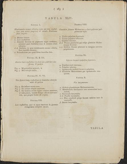
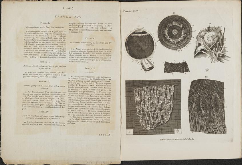
Tabula XLV illustrating cross-sections of the human eye and retina

Tabula XLV illustrating cross-sections of the human eye and retina, plus pages 263-264 of the anatomical treatise 'Schola et Historia Medicinae', by William Rowley, England, edition by Royce, England, 1851-1860

More

A human eye has been dissected in different ways to show the internal structure. The image in the top left shows the lens and the vitreous humour, the jelly-like substance that fills most of the eyeball. This print was taken from an anatomical treatise called 'Schola Medicinæ Universalis Nova' or the 'New Universal History and School of Medicine' by William Rowley (1742-1806), an English male midwife, surgeon and anatomist. First published in 1793, the work contained 68 copper engravings of the human body as well as a brief history of medicine. Anatomical prints were useful tools for medical students as specific features of the body could be enlarged and picked out, making the structures easier to understand.
Materials:
paper
Object Number:
1980-523/1/7
type:
print
Image ©
The Board of Trustees of the Science Museum
Tabula XLVI illustrating the muscles and nerves of the human eye

Tabula XLVI illustrating the muscles and nerves of the human eye

Tabula XLVI illustrating the muscles and nerves of the human eye, taken from the anatomical treatise 'Schola et Historia Medicinae', by William Rowley, England, edition by Royce, England, 1851-1860

More

This print shows the external muscles of the eye, which are responsible for moving the eyeball, viewed from different angles. This print was taken from an anatomical treatise called 'Schola Medicinæ Universalis Nova' or the 'New Universal History and School of Medicine' by William Rowley (1742-1806), an English male midwife, surgeon and anatomist. First published in 1793, the work contained 68 copper engravings of the human body as well as a brief history of medicine. Anatomical prints were useful tools for medical students as specific features of the body could be enlarged and picked out, making the structures easier to understand.

Materials:
paper
Object Number:
1980-523/1/8
type:
print