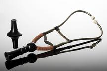
Curved array ultrasound transducer
- Made:
- 1980-1990 in United States and unknown place
Curved array ultrasound transducer, by ATL, 1980-1990 used for abdominal examination
Curved transducers are widely used to monitor pregnancy, and create the familiar cone-shape image of the inside of the womb. Since its invention in the late 1950s, ultrasound has been the technology of choice for observing unborn babies. Its popularity inspired technological development, so by the 1970s still images were replaced by moving ones, and by the 1990s, three-dimensional images were possible. This sort of transducer can also be used to look for cancers, cysts and blockages in the liver, kidney and pancreas.
Ultrasound was a transformative way to create cross-sectional images of inside the body, using high-frequency sound waves. This risk-free technique was pioneered in Glasgow by Professor Ian Donald, engineer Tom Brown and doctor John MacVicar in 1957. The first ultrasound images were recorded on black-and-white film and were always blurred. By the 1980s, technology had advanced to produce moving images with shades of grey, soon followed by 3D imaging.
Details
- Category:
- Clinical Diagnosis
- Object Number:
- 2023-490
- Measurements:
-
overall (estimated including cable: 40 mm x 800 mm x 850 mm,
- type:
- transducer