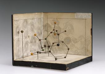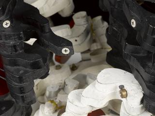
Icosahedron virus particle model made by Dr. June Almeida


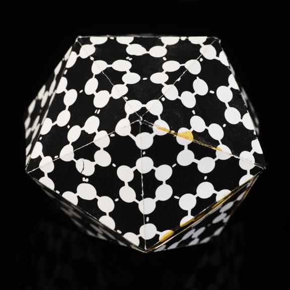
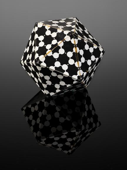
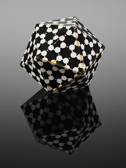
Icosahedron virus particle model, the second smallest in size of a set of four, made from black and white card, made by Dr. June Almeida (1930-2007), Virologist and pioneering electron microscopist, c.1960-1990.
June Almeida (née Hart) was an internationally renowned virologist who pioneered new electron microscopy methods for imaging and diagnosing viruses. June, with her colleagues, identified and named the first coronavirus in 1964, observing a round, grey dot covered in tiny spokes that formed a halo around the virus—like the sun’s corona. This virus model, made of card, was made by June Almeida for teaching. It shows the icosahedron shape of certain virus particles.
Born June Hart in 1930, she lived with her family in a tenement building in Glasgow, Scotland. At 16, she left school without funding to go to university and started working as a lab technician at Glasgow Royal Infirmary, where she used microscopes to help analyse tissue samples. She later emigrated to Canada, where she worked at the Ontario Cancer Institute in Toronto, developing new techniques in electron microscopy to image viruses. Amongst her scientific achievements was the first visualisation of the rubella virus, imaging hepatitis viruses, and developing the technique of antibody clumping to visualise common cold viruses. June finished her career at the Wellcome Research Laboratory, where she worked on developing diagnostic assays and vaccine development. She retired in 1985, where her career took a different direction as she qualified as a yoga teacher.
Details
- Category:
- Biochemistry
- Object Number:
- 2023-103/3
- Materials:
- paper (fibre product)
- Measurements:
-
overall (smallest): 36 mm x 55 mm x 55 mm,
overall (2nd Smallest): 55 mm x 70 mm x 70 mm,
overall (Largest): 75 mm x 100 mm x 100 mm,
overall (2nd Largest): 75 mm x 90 mm x 90 mm,
- type:
- virus model
