Electron microgram of magnesium oxide at high magnification (x 100,000) Electron microgram of magnesium oxide at high magnification
Electron micrograms of :- (a) Stalked bacteria, (b) Surface of wood, (c) Polystyrene latex, (d) Red ochre Electron micrograms
Electron micrograms of:- (a) 0.6% carbon steel - plastic replica, (b) 0.6% carbon steel - plastic replica shadowed with chromium Electron micrograms of:- (a) 0.6% carbon steel - plastic replica
Electron micrograms of:- (a) Surface of outer epicuticle of larva of tomato moth, (b) Potato virus X. Electron micrograms
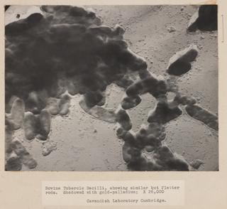
Electron micrograms of:- (a) Staphylococcal bacteriophage showing lysed coccus, (b) Bacteria showing flagellation, (c) Molecules of human haemoglobin, (d) Magnesium oxide crystals, showing crystalline interference bands, (e) Zinc oxide crystals, under dark field illumination, (f) Zinc oxide crystals, under light field illumination, (g) Staphylococcus and bacteriophage type 47B, showing coccus before attack by phage Electron micrograms of:- (a) Staphylococcal bacteriophage showing lysed coccus
Electron micrograms of:- (a) Silver halide crystals from the emulsion of a Kodak Nuclear Track plate, (b) Filamentary silver formed from single triangular silver halide crystals in a photographic emulsion during development, (c) Grains of Kodak Maximum Resolution plates, (d) A crystallised enzyme, orthozyme X. Electron micrograms
Electron micrograms of:- (a) Purified vaccinia virus, (b) Laked fowl red blood cell after absorption of influenza virus, showing only spherical virus particles, (c) Laked fowl red blood cell, (d) Laked fowl blood cell after absorption of influenza virus, showing both spherical and filamentous forms, (e) Same cell virus as in (d), by transmitted electron beam Electron micrograms of:- (a) Purified vaccinia virus