Microscope slide used during liquid-based cytology screening to deposit samples, made by Hologic, 2000-2017 Microscope slide used during cytology screening 2000-2017
Microscope slide containing human remains labelled ‘Path: Laboratory. 7826’, prepared by the Royal London Ophthalmic Hospital, London, England, 1837-1947 Microscope slide containing human remains labelled ‘Path: Laboratory. 7826’ 1837-1947
Prepared plastic slide belonging to the Tasco 300x children's microscope, made in China for Tasco, United States, 1996 Prepared plastic slide belonging to the Tasco 300x children's microscope 1996
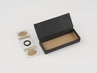
Microscope slide with 28 x 28mm collodion reproduction of grid numbered 1-50 on each axis - squares marked 1/1-50/50. Maltwood finder and manufactured name inscribed diamond. R. & J. beck. R. & J. Beck maltwood finder 1853-1863
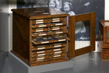
Case containing 24 microscope slides comprised of human remains demonstrating pathological conditions labelled ‘Prize Medal, Paris, 1867’, prepared by Arthur C. Cole and Son, England, 1867 Case containing 24 microscope slides comprised of human remains demonstrating pathological conditions 1867
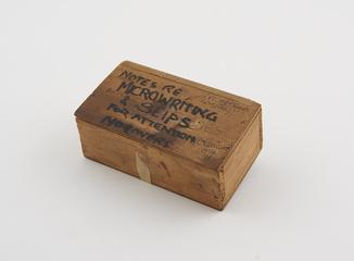
Box of microscope slides belonging to J H Fisher, a surgeon at the Royal London Ophthalmic Hospital (now Moorfield Eye Hospital) Box of microscope slides belonging to J H Fisher 1867-1933
Box containing 12 microscope slides comprised of human remains, prepared by Edmund Wheeler, Holloway, London, 1860-1884 Box containing 12 microscope slides comprised of human remains 1860-1884
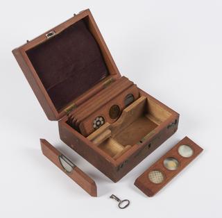
Eight microscope slides collected by Professor P. A. Buxton from the London School of Hygiene & Tropical Medicine, each showing parastic larvae, in wooden box Microscope slides showing parastic larvae