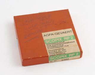
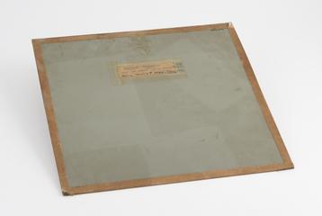
Scopix RP 2 fluorographic film, part of mass miniature radiography set, Europe, 1960-1961
1960-1961
1960-1961
c.1965
1960-1961

1960-1961

1930-1939

1896-1900

1890-1900
1896-01-20
1987
1960-1961
1960-1961

1960-1961

1896-01-20
1896
circa 1902
1905-1909
1915
1905-1909
1915