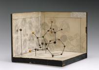


![]() This image is released under a CC BY-NC-SA 4.0 Licence
This image is released under a CC BY-NC-SA 4.0 Licence
Buy this image as a print
BuyLicense this image for commercial use at Science and Society Picture Library
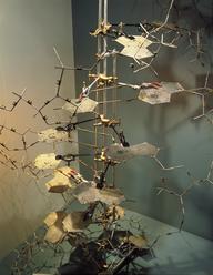
License![]() This image is released under a CC BY-NC-SA 4.0 Licence
This image is released under a CC BY-NC-SA 4.0 Licence
Buy this image as a print
BuyLicense this image for commercial use at Science and Society Picture Library
License![]() This image is released under a CC BY-NC-SA 4.0 Licence
This image is released under a CC BY-NC-SA 4.0 Licence
Buy this image as a print
BuyLicense this image for commercial use at Science and Society Picture Library
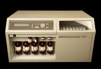
LicenseCobas bio centrifugal analyser.
Detail view .
Science Museum Group Collection
© The Board of Trustees of the Science Museum
Cobas bio centrifugal analyser
Science Museum Group Collection
© The Board of Trustees of the Science Museum
Cobas bio centrifugal analyser
Science Museum Group Collection
© The Board of Trustees of the Science Museum
COBAS® bio centrifugal analyser, Roche Diagnostics, Switzerland, 1988-1998
Introduced in 1979 by Roche Diagnostics this machine analyses blood. It contains a centrifugal analyser that can handle seven different clinical analysis procedures. To analyse a sample of blood is put into a special cup. The cup is placed in a tray in the machine and its location detected by a light beam. A needle pierces the cup lid and transfers a very small amount of the sample to the small vertical compartment on the rotor on the right. Another probe moves reagent from the sample disc on the left to the larger vertical compartment on the rotor. The rotor has 30 clear acrylic cuvettes (small compartments) arranged in a circle. When the centrifuge spins the rotor, the sample and reagent combine in the reaction cuvette. A high-intensity light beam produced by a xenon flash tube then analyses the mix through a grating spectrophotometer. The analysis results this can be printed out or transferred by electronic interface to a computer. This is machine was used in the Pathology unit at Grimsby Hospital from 1979 until the late 1990s.
Details
- Category:
- Biochemistry
- Object Number:
- 1998-875
- Materials:
- glass, metal (unknown) and plastic (unidentified)
- Measurements:
-
overall: 825 mm x 890 mm x 510 mm, 140kg
- type:
- centrifugal analyser
- credit:
- Department of Pathology, Grimsby General Hospital