Anatomical model of the spine
- maker:
- Louis Thomas Jerome Auzoux


![]() This image is released under a CC BY-NC-SA 4.0
Licence
This image is released under a CC BY-NC-SA 4.0
Licence
License this image for commercial use at Science and Society Picture Library
License![]() This image is released under a CC BY-NC-SA 4.0
Licence
This image is released under a CC BY-NC-SA 4.0
Licence
License this image for commercial use at Science and Society Picture Library
License![]() This image is released under a CC BY-NC-SA 4.0
Licence
This image is released under a CC BY-NC-SA 4.0
Licence
License this image for commercial use at Science and Society Picture Library
License![]() This image is released under a CC BY-NC-SA 4.0
Licence
This image is released under a CC BY-NC-SA 4.0
Licence
License this image for commercial use at Science and Society Picture Library
LicenseAnatomical model of the spine, papier mache, in two parts
Science Museum Group Collection
© The Board of Trustees of the Science Museum
Anatomical model of the spine, papier mache, in two parts
Science Museum Group Collection
© The Board of Trustees of the Science Museum
Anatomical model of the spine, papier mache, in two parts
Science Museum Group Collection
© The Board of Trustees of the Science Museum
Anatomical model of the spine, papier mache, in two parts
Science Museum Group Collection
© The Board of Trustees of the Science Museum
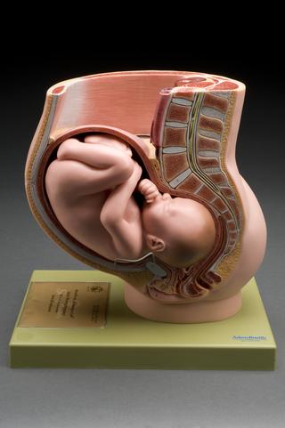
Anatomical model of the spine, papier mâché, in two parts, labelled, by Dr. Auzoux, French, 1901.
Made from papier mâché, this anatomical model shows the internal structure of the human spine, including the spinal cord which, with the brain, makes up the central nervous system. The model was made by the factory of Louis Thomas Jérôme Auzoux (1797-1880), a French physician and model maker who developed a large business producing anatomical models. The spine was probably used when teaching students anatomy. Models can be used to emphasise and enlarge minute anatomical structures, making them easier to understand, and unlike human tissue they do not need to be preserved.