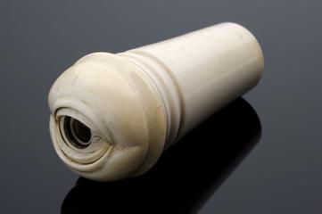
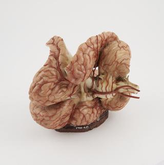
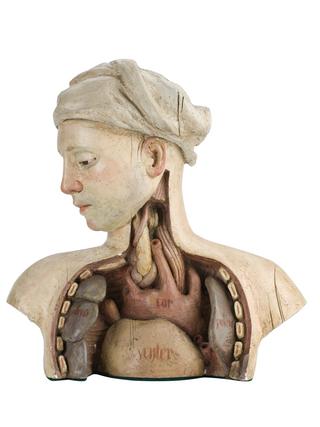
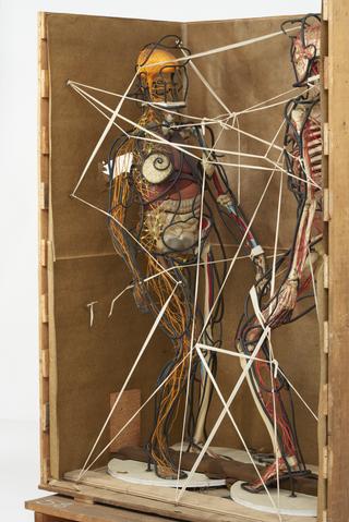
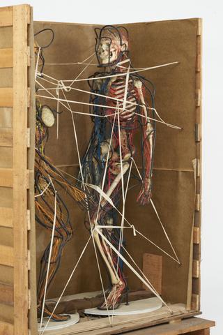
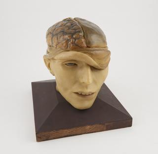
Wax anatomical model of a female showing internal organs, Florence, Italy, 1818 Anatomy & Pathology 1818
Wax anatomical model of female torso and head, showing internal organs, with removable heart, on wooden display cabinet, by Francesco Calenzuoli, Italian, dated 1818 Wax anatomical model of a female showing internal organs, Florence, Italy, 1818 Anatomy & Pathology 1818
Plasticene model of human uterus at eleven weeks gestation Obstetrics, Gynaecology & Contraception 1941
Anatomical female figure. A small ivory model with removable anterior thoracic and abdominal wall Anatomy & Pathology
Anatomical model of the eye with a descriptive booklet in a leather case, Italian, 1674-1679 Ophthalmology 1674-1679
Plasticene model of the human uterine blood supply mounted in box Obstetrics, Gynaecology & Contraception 1941
Wax fragment found underneath legs of wooden display board, Italian, dated 1818 Part of wax anatomical model of a female showing internal organs, Florence, Italy, 1818 Anatomy & Pathology 1818
Wax fragment from anatomical model of female torso and head, showing internal organs, by Francesco Calenzuoli, Italian, dated 1818 Part of wax anatomical model of a female showing internal organs, Florence, Italy, 1818 Anatomy & Pathology 1818
Silk fragments from Wax anatomical model of female torso and head, showing internal organs, with removable heart, on wooden display cabinet, by Francesco Calenzuoli, Italian, 1818 Part of wax anatomical model of a female showing internal organs, Florence, Italy, 1818 Anatomy & Pathology 1818
Fragment of resin (found by hip area on proper left hand side), from wax anatomical model of female torso and head, showing internal organs, by Francesco Calenzuoli, Italian, dated 1818 Part of wax anatomical model of a female showing internal organs, Florence, Italy, 1818 Anatomy & Pathology 1818
Fragments from back of wax anatomical model of female torso and head, showing internal organs, with removable heart, by Francesco Calenzuoli, Italian, dated 1818 Part of wax anatomical model of a female showing internal organs, Florence, Italy, 1818 Anatomy & Pathology 1818
Fragment of wax and hair, found underneath head of wax anatomical model of female torso and head, showing internal organs, with removable heart, on wooden display cabinet, by Francesco Calenzuoli, Italian, dated 1818 Part of wax anatomical model of a female showing internal organs, Florence, Italy, 1818 Anatomy & Pathology 1818
Wax fragment from removable heart from wax anatomical model of female torso and head, showing internal organs by Francesco Calenzuoli, Italian, dated 1818 Part of wax anatomical model of a female showing internal organs, Florence, Italy, 1818 Anatomy & Pathology 1818
The Ho model eye demonstrating basic principles of vision. With a brass base and pillar supporting tube fitted with the iris diaphragm. There is a provision for inserting glass lenses, ground on rear surface, and different curvatures on front face. The lenses are marked I, II, III, and IIII. F.E. Becker and Co. c.1913 The 'Ho Model' eye Photographic Technology 1909-1919
Full-size female animated figure model, part of installation with motorised base and four hear phones through which recording of narration about the human body can be played, from Cleveland Health museum, Ohio, United States, 1965 Female animated figure model from Cleveland Health Museum, Ohio, United States, 1965 Anatomy & Pathology 1965