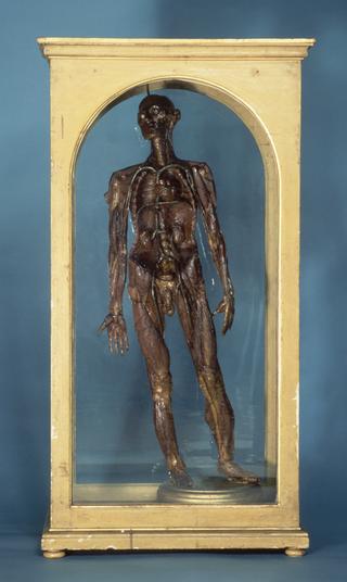
Wax anatomical model of a female showing internal organs, Florence, Italy, 1818

Wax anatomical model of female torso and head Wax anatomical model of female torso and head Wax anatomical model of female torso and head Wax anatomical model of female torso and head Wax anatomical model of female torso and head Wax anatomical model of female torso and head Wax anatomical model of female torso and head Wax anatomical model of female torso and head Wax anatomical model of female torso and head Wax anatomical model of female torso and head Wax anatomical model of a female showing internal organs, Florence, Italy, 1818 Wax anatomical model of a female showing internal organs, Florence, Italy, 1818 Wax anatomical model of a female showing internal organs, Florence, Italy, 1818 Wax anatomical model of female torso and head
Buy this image as a print 

Buy

License this image for commercial use at Science and Society Picture Library

License

Buy this image as a print 

Buy

License this image for commercial use at Science and Society Picture Library

License

Buy this image as a print 

Buy

License this image for commercial use at Science and Society Picture Library

License

Buy this image as a print 

Buy

License this image for commercial use at Science and Society Picture Library

License

Buy this image as a print 

Buy

License this image for commercial use at Science and Society Picture Library

License

Buy this image as a print 

Buy

License this image for commercial use at Science and Society Picture Library

License

Buy this image as a print 

Buy

License this image for commercial use at Science and Society Picture Library

License

Creative Commons LicenseThis image is released under a CC BY-NC-SA 4.0 Licence

Buy this image as a print 

Buy

License this image for commercial use at Science and Society Picture Library

License

Creative Commons LicenseThis image is released under a CC BY-NC-SA 4.0 Licence

Buy this image as a print 

Buy

License this image for commercial use at Science and Society Picture Library

License

Creative Commons LicenseThis image is released under a CC BY-NC-SA 4.0 Licence

Buy this image as a print 

Buy

License this image for commercial use at Science and Society Picture Library

License

Creative Commons LicenseThis image is released under a CC BY-NC-SA 4.0 Licence

Buy this image as a print 

Buy

License this image for commercial use at Science and Society Picture Library

License

Creative Commons LicenseThis image is released under a CC BY-NC-SA 4.0 Licence

Buy this image as a print 

Buy

License this image for commercial use at Science and Society Picture Library

License

Creative Commons LicenseThis image is released under a CC BY-NC-SA 4.0 Licence

Buy this image as a print 

Buy

License this image for commercial use at Science and Society Picture Library

License

Creative Commons LicenseThis image is released under a CC BY-NC-SA 4.0 Licence

Buy this image as a print 

Buy

License this image for commercial use at Science and Society Picture Library

License

Wax anatomical model of female torso and head
Science Museum Group
© The Board of Trustees of the Science Museum, London.

Wax anatomical model of female torso and head
Science Museum Group
© The Board of Trustees of the Science Museum, London.

Wax anatomical model of female torso and head
Science Museum Group
© The Board of Trustees of the Science Museum, London

Wax anatomical model of female torso and head
Science Museum Group
© The Board of Trustees of the Science Museum, London.

Wax anatomical model of female torso and head
Science Museum Group
© The Board of Trustees of the Science Museum, London.

Wax anatomical model of female torso and head
Science Museum Group
© The Board of Trustees of the Science Museum, London.

Wax anatomical model of female torso and head
Science Museum Group
© The Board of Trustees of the Science Museum, London.

Wax anatomical model of female torso and head
Science Museum Group Collection
© The Board of Trustees of the Science Museum

Wax anatomical model of female torso and head
Science Museum Group Collection
© The Board of Trustees of the Science Museum

Wax anatomical model of female torso and head
Science Museum Group Collection
© The Board of Trustees of the Science Museum

Science Museum Group Collection
© The Board of Trustees of the Science Museum

Science Museum Group Collection
© The Board of Trustees of the Science Museum

Science Museum Group Collection
© The Board of Trustees of the Science Museum

Wax anatomical model of female torso and head
Science Museum Group Collection
© The Board of Trustees of the Science Museum

Wax anatomical model of female torso and head, showing internal organs, with removable heart, on wooden display cabinet, by Francesco Calenzuoli, Italian, dated 1818

This anatomical wax model shows the internal organs in a female torso and head, including the lungs, liver, stomach, kidneys and intestines. Complete with the veins and arteries, the heart is entirely removable.

The figure was made by Francesco Calenzuoli (1796-1821), an Italian model maker renowned for his attention to detail. Wax models were used for teaching anatomy to medical students because they made it possible to pick out and emphasise specific features of the body, making their structure and function easier to understand. This made them especially useful at a time when few bodies were available for dissection. The model was donated by the Department of Human Anatomy at the University of Oxford.

Details

Category:
Anatomy & Pathology
Object Number:
1988-249
Materials:
model, wax, model, silk, model, hair, cabinet, wood and cabinet, glass
Measurements:
overall: 20 kg
type:
anatomical model
credit:
Oxford University (Dept. Human Anatomy)

Parts

Wax anatomical model of a female showing internal organs, Florence, Italy, 1818

Wax anatomical model of a female showing internal organs, Florence, Italy, 1818

Wax anatomical model of female torso and head, showing internal organs, with removable heart, on wooden display cabinet, by Francesco Calenzuoli, Italian, dated 1818

More

This anatomical wax model shows the internal organs in a female torso and head, including the lungs, liver, stomach, kidneys and intestines. Complete with the veins and arteries, the heart is entirely removable.

The figure was made by Francesco Calenzuoli (1796-1821), an Italian model maker renowned for his attention to detail. Wax models were used for teaching anatomy to medical students because they made it possible to pick out and emphasise specific features of the body, making their structure and function easier to understand. This made them especially useful at a time when few bodies were available for dissection. The model was donated by the Department of Human Anatomy at the University of Oxford.

Part of wax anatomical model of a female showing internal organs, Florence, Italy, 1818

Part of wax anatomical model of a female showing internal organs, Florence, Italy, 1818

Fragment of resin (found by hip area on proper left hand side), from wax anatomical model of female torso and head, showing internal organs, by Francesco Calenzuoli, Italian, dated 1818

More

This anatomical wax model shows the internal organs in a female torso and head, including the lungs, liver, stomach, kidneys and intestines. Complete with the veins and arteries, the heart is entirely removable.

The figure was made by Francesco Calenzuoli (1796-1821), an Italian model maker renowned for his attention to detail. Wax models were used for teaching anatomy to medical students because they made it possible to pick out and emphasise specific features of the body, making their structure and function easier to understand. This made them especially useful at a time when few bodies were available for dissection. The model was donated by the Department of Human Anatomy at the University of Oxford.

Part of wax anatomical model of a female showing internal organs, Florence, Italy, 1818

Part of wax anatomical model of a female showing internal organs, Florence, Italy, 1818

Wax fragment from removable heart from wax anatomical model of female torso and head, showing internal organs by Francesco Calenzuoli, Italian, dated 1818

More

This anatomical wax model shows the internal organs in a female torso and head, including the lungs, liver, stomach, kidneys and intestines. Complete with the veins and arteries, the heart is entirely removable.

The figure was made by Francesco Calenzuoli (1796-1821), an Italian model maker renowned for his attention to detail. Wax models were used for teaching anatomy to medical students because they made it possible to pick out and emphasise specific features of the body, making their structure and function easier to understand. This made them especially useful at a time when few bodies were available for dissection. The model was donated by the Department of Human Anatomy at the University of Oxford.

Part of wax anatomical model of a female showing internal organs, Florence, Italy, 1818

Part of wax anatomical model of a female showing internal organs, Florence, Italy, 1818

Wax fragment from anatomical model of female torso and head, showing internal organs, by Francesco Calenzuoli, Italian, dated 1818

More

This anatomical wax model shows the internal organs in a female torso and head, including the lungs, liver, stomach, kidneys and intestines. Complete with the veins and arteries, the heart is entirely removable.

The figure was made by Francesco Calenzuoli (1796-1821), an Italian model maker renowned for his attention to detail. Wax models were used for teaching anatomy to medical students because they made it possible to pick out and emphasise specific features of the body, making their structure and function easier to understand. This made them especially useful at a time when few bodies were available for dissection. The model was donated by the Department of Human Anatomy at the University of Oxford.

Part of wax anatomical model of a female showing internal organs, Florence, Italy, 1818

Part of wax anatomical model of a female showing internal organs, Florence, Italy, 1818

Silk fragments from wax anatomical model of female torso and head, showing internal organs, with removable heart, on wooden display cabinet, by Francesco Calenzuoli, Italian, 1818

More

This anatomical wax model shows the internal organs in a female torso and head, including the lungs, liver, stomach, kidneys and intestines. Complete with the veins and arteries, the heart is entirely removable.

The figure was made by Francesco Calenzuoli (1796-1821), an Italian model maker renowned for his attention to detail. Wax models were used for teaching anatomy to medical students because they made it possible to pick out and emphasise specific features of the body, making their structure and function easier to understand. This made them especially useful at a time when few bodies were available for dissection. The model was donated by the Department of Human Anatomy at the University of Oxford.

Materials:
wax
Object Number:
1988-249/5
type:
anatomical model
Part of wax anatomical model of a female showing internal organs, Florence, Italy, 1818

Part of wax anatomical model of a female showing internal organs, Florence, Italy, 1818

Fragments from back of wax anatomical model of female torso and head, showing internal organs, with removable heart, by Francesco Calenzuoli, Italian, dated 1818

More

This anatomical wax model shows the internal organs in a female torso and head, including the lungs, liver, stomach, kidneys and intestines. Complete with the veins and arteries, the heart is entirely removable.

The figure was made by Francesco Calenzuoli (1796-1821), an Italian model maker renowned for his attention to detail. Wax models were used for teaching anatomy to medical students because they made it possible to pick out and emphasise specific features of the body, making their structure and function easier to understand. This made them especially useful at a time when few bodies were available for dissection. The model was donated by the Department of Human Anatomy at the University of Oxford.

Object Number:
1988-249/6
type:
anatomical model
Part of wax anatomical model of a female showing internal organs, Florence, Italy, 1818

Part of wax anatomical model of a female showing internal organs, Florence, Italy, 1818

Wax fragment found underneath legs of wooden display board, Italian, dated 1818

More

This anatomical wax model shows the internal organs in a female torso and head, including the lungs, liver, stomach, kidneys and intestines. Complete with the veins and arteries, the heart is entirely removable.

The figure was made by Francesco Calenzuoli (1796-1821), an Italian model maker renowned for his attention to detail. Wax models were used for teaching anatomy to medical students because they made it possible to pick out and emphasise specific features of the body, making their structure and function easier to understand. This made them especially useful at a time when few bodies were available for dissection. The model was donated by the Department of Human Anatomy at the University of Oxford.

Materials:
wax
Object Number:
1988-249/7
type:
anatomical model
Part of wax anatomical model of a female showing internal organs, Florence, Italy, 1818

Part of wax anatomical model of a female showing internal organs, Florence, Italy, 1818

Fragment of wax and hair, found underneath head of wax anatomical model of female torso and head, showing internal organs, with removable heart, on wooden display cabinet, by Francesco Calenzuoli, Italian, dated 1818

More

This anatomical wax model shows the internal organs in a female torso and head, including the lungs, liver, stomach, kidneys and intestines. Complete with the veins and arteries, the heart is entirely removable.

The figure was made by Francesco Calenzuoli (1796-1821), an Italian model maker renowned for his attention to detail. Wax models were used for teaching anatomy to medical students because they made it possible to pick out and emphasise specific features of the body, making their structure and function easier to understand. This made them especially useful at a time when few bodies were available for dissection. The model was donated by the Department of Human Anatomy at the University of Oxford.