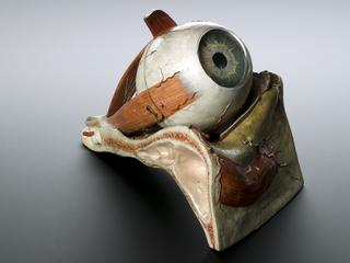
Wax anatomical model of thorax showing ecthyma luridum

Wax anatomical model of thorax showing ecthyma luridum Wax anatomical model of thorax showing ecthyma luridum Wax anatomical model of thorax showing ecthyma luridum Wax anatomical model of thorax showing ecthyma luridum Wax anatomical model of thorax showing ecthyma luridum

Creative Commons LicenseThis image is released under a CC BY-NC-SA 4.0 Licence

Buy this image as a print 

Buy

License this image for commercial use at Science and Society Picture Library

License

Creative Commons LicenseThis image is released under a CC BY-NC-SA 4.0 Licence

Buy this image as a print 

Buy

License this image for commercial use at Science and Society Picture Library

License

Creative Commons LicenseThis image is released under a CC BY-NC-SA 4.0 Licence

Buy this image as a print 

Buy

License this image for commercial use at Science and Society Picture Library

License

Creative Commons LicenseThis image is released under a CC BY-NC-SA 4.0 Licence

Buy this image as a print 

Buy

License this image for commercial use at Science and Society Picture Library

License

Creative Commons LicenseThis image is released under a CC BY-NC-SA 4.0 Licence

Buy this image as a print 

Buy

License this image for commercial use at Science and Society Picture Library

License

Wax anatomical model of thorax showing ecthyma luridum
Science Museum Group
© The Board of Trustees of the Science Museum, London.

Wax anatomical model of thorax showing ecthyma luridum
Science Museum Group
© The Board of Trustees of the Science Museum, London.

Wax anatomical model of thorax showing ecthyma luridum
Science Museum Group
© The Board of Trustees of the Science Museum, London.

Wax anatomical model of thorax showing ecthyma luridum
Science Museum Group
© The Board of Trustees of the Science Museum, London.

Wax anatomical model of thorax showing ecthyma luridum
Science Museum Group Collection
© The Board of Trustees of the Science Museum

Wax anatomical model of thorax showing ecthyma luridum, in glass case, by Joseph Towne, English, 1825-1879

Wax anatomical models such as this were for teaching purposes. They were created by skilled craftsman and had to be realistic. This example was almost certainly modelled on a dead body because the word ‘autopsy’ features on the label. This example shows the thorax, or chest area, covered in pus-filled boils caused by the skin disease ecthyma luridum. This causes inflammation and spots to form on the skin. During the 1830s, physicians believed it was associated with people with ‘broken constitutions’ and was treatable with warm sulphurous baths. It is now easily treated with medicated creams.

Joseph Towne was a wax modeller for Guy's Hospital, London for over 50 years, He completed several hundred models. In 1826, aged just 20 years-old he submitted his first model to the Royal Society of Arts and was awarded a silver medal. He won a gold medal from the same institution in 1827. Many of his models were based on direct observation of the human body via autopsy specimens. They are still useful teaching resources.

Details

Category:
Anatomy & Pathology
Object Number:
1986-455
Materials:
model, wax and case, glass
Measurements:
overall: 304 mm x 244 mm x 186 mm,
type:
anatomical model
credit:
Bridge City Auction Service